


UV-Vis Spectroscopy for Characterising the Optical Properties of Gold Nanoparticles
- UV-Vis spectroscopy is one of the best and easiest techniques for characterising Noble metal nanoparticles, such as gold.
- Gold nanoparticles are useful for colourimetric and optical sensing applications because their optical properties are based on localised surface plasmon resonance (LSPR).
- LSPR is highly tuneable and changes in response to external factors that change particle size, morphology, interparticle distance, or the refractive index of the surrounding medium.
- LSPR is seen on a UV-Vis spectrum as a distinct peak. Changes in peak wavelength or shape are easily detectable.
Introduction
The use of gold nanoparticles (AuNPs) predates the scientific revolution, with the earliest known use being in the stained glass of the Lycurgus cup, thought to be preserved from the fourth century, A.D.2 They have also been used frequently in stained glass windows, Figure 1. It is understood that gold particles dispersed in the glass are the cause of the deep red colour observable when light is shone through it. A nanoparticle (NP) is defined as a particle with at least one dimension between 1 and 100 nm in size. Metallic NPs have become abundant in scientific research because they exhibit interesting and tuneable physical, optical, and chemical properties when compared with their bulk metal counterparts. This is true of AuNPs in particular, which have optical properties that are easily manipulated and related to their physical state and chemical environment.1 This means that they can be precisely engineered for a wide range of different applications.

Figure 1: Gold nanoparticles are responsible for the red tint in stained glass windows and pieces of art.
Today, colloidal AuNPs are particularly useful for colourimetric and optical detection applications because they are efficient at absorbing and scattering light and can be tuned to exhibit optical activity at different wavelengths based on their physical and chemical characteristics. More specifically, they are capable of supporting a localised surface plasmon resonance (LSPR), caused by a displacement and coherent oscillation of the conduction electrons on the NP surface at a particular frequency when excited by incident light.3 The LSPR is an extremely useful parameter for optical sensing because its frequency depends strongly on several factors including NP size, NP morphology, interparticle distance, and the refractive index of the surrounding dielectric medium, which can all be designed to change in response to an external variable in a detection assay. The optical properties of AuNPs are traditionally probed using UV-Vis spectroscopy because it is an excellent technique for characterising a sample’s response to light over a large wavelength range and the LSPR appears on the absorption spectrum of AuNPs as a distinct peak.4 Hence, in this Application note, the optical properties of AuNPs are investigated using UV-Vis spectroscopy. Firstly, the LSPR of spherical colloidal AuNPs is investigated as a function of particle diameter. Then, the chemical sensing potential of AuNPs is demonstrated by tracking the LSPR during an induced aggregation experiment.
Materials and Methods
UV-Vis spectroscopic measurements were performed on Edinburgh Instruments DS5 UV-Vis Spectrophotometer. Gold nanoparticle colloid solutions of various sizes were purchased from BBI Solutions. Sodium chloride was purchased from Sigma Aldrich. To induce aggregation in the AuNPs, sodium chloride was dissolved in distilled water and a small aliquot was then added to a solution of AuNPs. For UV-Vis absorption measurements, a 1.5 mL aliquot of the sample was placed in a 1 cm path length cuvette in the sample light path and distilled water was placed in a cuvette in the reference light path. Samples were analysed between 400 nm and 1000 nm with a scan speed of 800 nm/min and a read interval of 1 nm.
Results and Discussion
To investigate the relationship between the particle diameter and the LSPR, various spherical AuNP samples with particle diameters between 10 and 80 nm were characterised using UV-Vis spectroscopy, Figure 2. Spherical AuNPs that are stable in solution are easily identifiable in UV-Vis spectroscopy because they exhibit a sharp absorption band, with a λmax normally between 500 nm and 600 nm, as is shown in Figure 2. The data also shows that as the size of the NPs increases, the LSPR wavelength, based on the λmax values in the normalised absorbance spectra, redshifts. This change of the LSPR to a longer wavelength is a result of the electrons oscillating over a larger area and hence at a lower frequency. The LSPR redshift with increasing particle size is also useful in the design of optical nanosensors for biological purposes because such applications require near-infrared (NIR) excitation sources. A colour change is also seen with increasing particle size, and this is explained by the corresponding absorption spectra. The smaller NPs absorb strongly in the green portion of the visible spectrum, resulting in a rich ruby-red colour that makes them ideal for colourimetric assays such as lateral flow testing devices. As the NP size increases and the LSPR redshifts, more red light is absorbed and the colloidal solution tends to exhibit a purple hue.
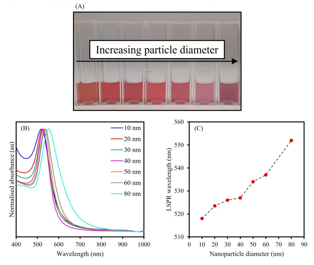
Figure 2. The effect of increasing particle diameter on the LSPR of spherical AuNPs.
UV-Vis spectroscopy, and by extension the LSPR, can also be used to track changes in the stability of colloidal AuNPs, the loss of which results in the aggregation of individual NPs into clusters that alters their size, shape, and interparticle distance. When aggregation is induced in a controlled manner, it is a useful property that can be used in colourimetric and LSPR assays, and in designing nanosensors for NIR biological applications.5 However, when not controlled it can also cause the irreversible dissolution of NPs.
To demonstrate the effect of aggregation on LSPR, 20 nm AuNPs were mixed with salt solutions of varying concentrations and analysed using UV-Vis spectroscopy, Figure 3. AuNPs are traditionally prepared using a technique that involves the reduction of a gold salt with sodium citrate, and therefore the particles are capped by citrate anions. The net negative charge on the surface of each AuNP contributes heavily to the stability of the colloid. When salt is added this negative charge is masked, and the electrostatic interactions that initially kept the AuNPs from aggregating are removed, which reduces the stability of the colloid dramatically. In the absence of an aggregating agent, the 20 nm AuNPs exhibit an LSPR at 524 nm and are a deep red colour. When a low concentration of the salt solution, intended to induce a controlled aggregation, is mixed with the NPs, they become pink in colour but remain stable in the colloid. The absorbance spectrum of this solution also shows distinct changes from the bare AuNPs. Even though the λmax remains consistent with the bare NPs, the absorbance of the LSPR peak is dampened and there is an increased response in the NIR range between 600 nm and 1000 nm. The dampening of the original LSPR band shows that the number of stable 20 nm NPs in the colloid has been depleted as a result of aggregation. Further, the increased optical activity in the NIR range is suggestive of aggregation because it indicates that the free electrons on the surface of each NP have become delocalised and are being shared among other NPs within aggregated clusters, thus making them resonate at a lower frequency than the original LSPR. When a larger concentration of salt is mixed with the bare AuNPs, these effects are even more pronounced, and the solution becomes purple in colour before the aggregates come out of the solution. Moreover, the λmax of the original LSPR peak red-shifts from 524 nm to 532 nm, which again shows the delocalisation of surface electrons as a result of NP clusters forming. The susceptibility of citrate-capped AuNPs to salt-induced aggregation can become a problem especially when the AuNPs are introduced to biological media; therefore, it is common for them to be encapsulated beforehand in silica or polyethylene glycol which increases their stability and biocompatibility.
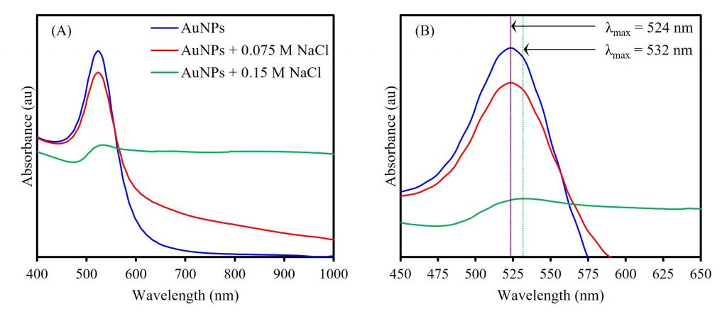
Figure 3. UV-Vis absorption spectra (A) showing salt-induced aggregation’s effect on AuNP LSPR and (B) comparing LSPR of the bare AuNP, controlled AuNP aggregate, and uncontrolled AuNP aggregate solutions.
Conclusion
In this Application Note, the Edinburgh Instruments DS5 Dual Beam UV-Vis Spectrophotometer was used to characterise the optical properties of various AuNP samples. Gold nanoparticles have become incredibly popular in a wide range of sensing applications, and UV-Vis spectroscopy is an excellent technique for ensuring the stability of samples and fine-tuning their optical, physical, and chemical properties.
References
- R. Sardar et al., Gold Nanoparticles: Past, Present, and Future, Langmuir, 2009, 25, 13840–13851.
- The British Museum, Drinking-Cup, https://www.britishmuseum.org/collection/object/H_1958-1202-1 (accessed January 2023).
- A. Willets et al., Localized Surface Plasmon Resonance Spectroscopy and Sensing, Annu. Rev. Phys. Chem., 2007, 58, 267–297.
- W. Haiss et al., Determination of Size and Concentration of Gold Nanoparticles from UV-Vis Spectra, Anal. Chem., 2007, 79, 4215–4221.
- S. Unser et al., Localized Surface Plasmon Resonance Biosensing: Current Challenges and Approaches, Sensors (Basel), 2015, 15, 15684–15716
