


Feature Highlight: MicroPL Upgrade
The MicroPL Upgrade for the FLS1000 and FS5 photoluminescence spectrometers enables widefield imaging, single point spectra and lifetime, and lifetime imaging of microscopic photoluminescent samples.
Widefield Imaging
In widefield imaging, excitation light from the spectrometer excitation monochromator is used to illuminate an area of the sample. The monochromator can either be set to zero order to generate white light for brightfield imaging or to a specific excitation wavelength for photoluminescence widefield imaging. Reflected light or photoluminescence from the sample is then detected using the widefield imaging camera. The widefield photoluminescence image of phosphor microparticles is shown in Figure 2.
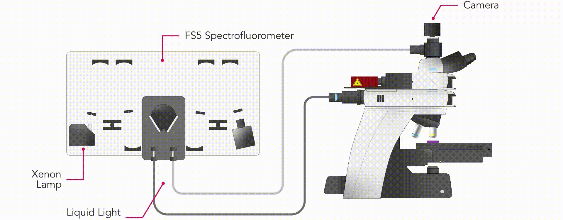
Figure 1: MicroPL configuration for widefield imaging.
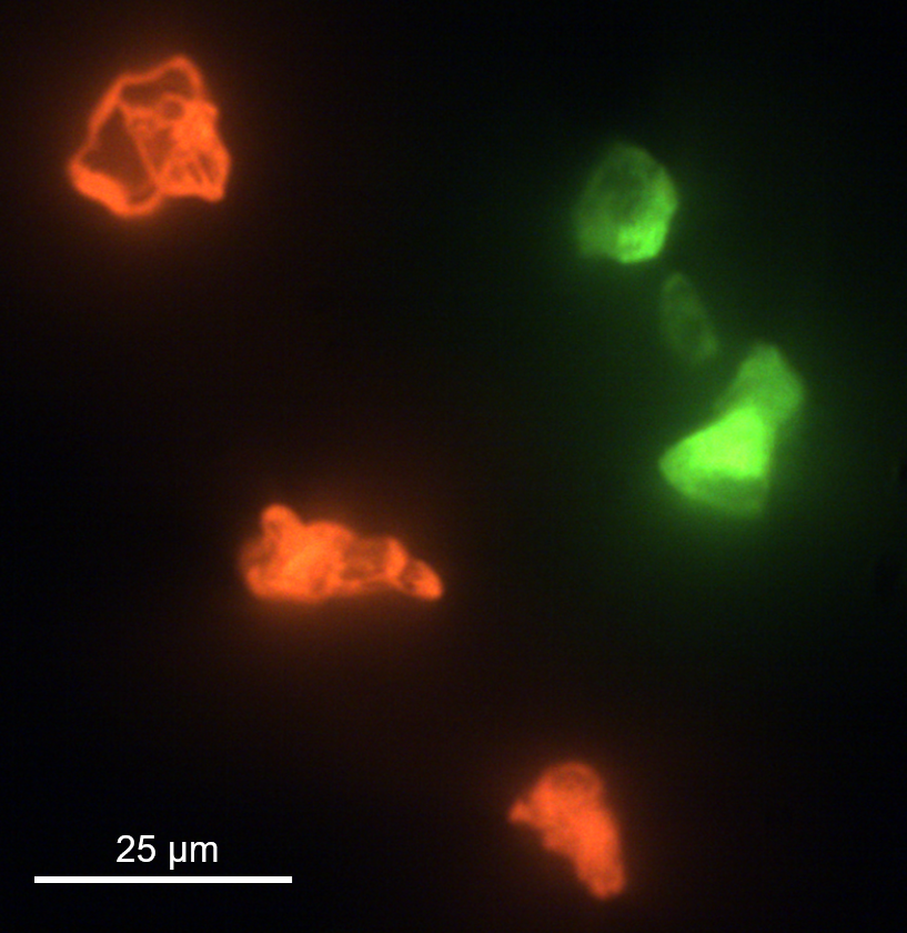
Figure 2: Widefield photoluminescence of phosphor microparticles.
Single Point Spectra & Lifetime
After acquiring the widefield image, the properties of the sample can be analysed in more detail using single-point excitation. In point excitation, an EPL, HPL or VPL laser is used for excitation which can provide an excitation spot size down to a few microns. The photoluminescence from the sample is directed back to the spectrometer where it is wavelength selected and detected. Using single-point excitation the photoluminescence spectra and lifetime of the phosphor microparticles were measured (Figure 4).
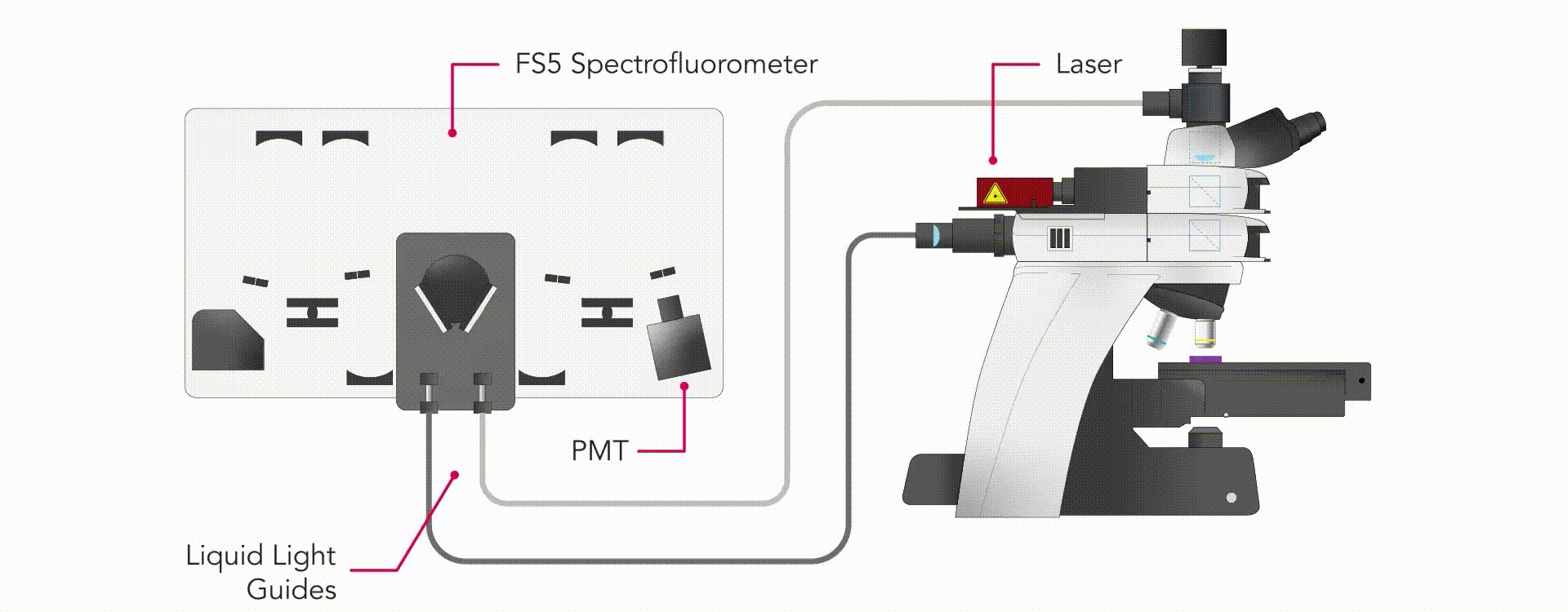
Figure 3: MicroPL configuration for single point spectra & lifetime.
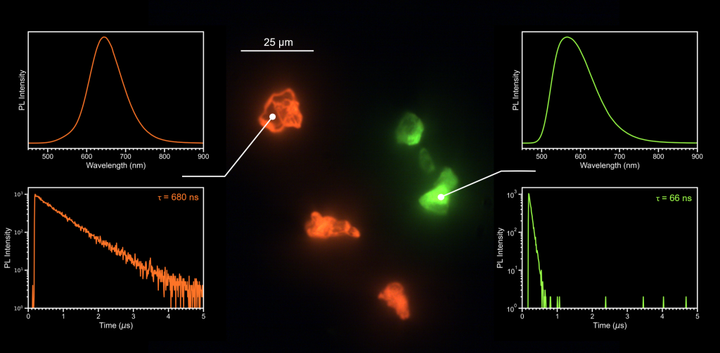
Figure 4: Photoluminescence spectra and lifetimes of two of the phosphor microparticles.
Lifetime Mapping
The motorised stage of the MicroPL upgrade enables the photoluminescence lifetime across the sample surface to be mapped. In lifetime mapping the photoluminescence decay at each point in the grid is acquired to create a lifetime image of the sample surface. Lifetime maps can be acquired using either time-correlated single photon counting (TCSPC) or multichannel scaling (MCS) with Edinburgh Instruments EPL, HPL and VPL pulsed diode lasers for excitation. Using lifetime mapping the phosphor microparticles can be distinguished by their lifetime, with the particles coloured in red having a lifetimee of 680 ns compared to 66 ns of the particles shown in blue (Figure 6).
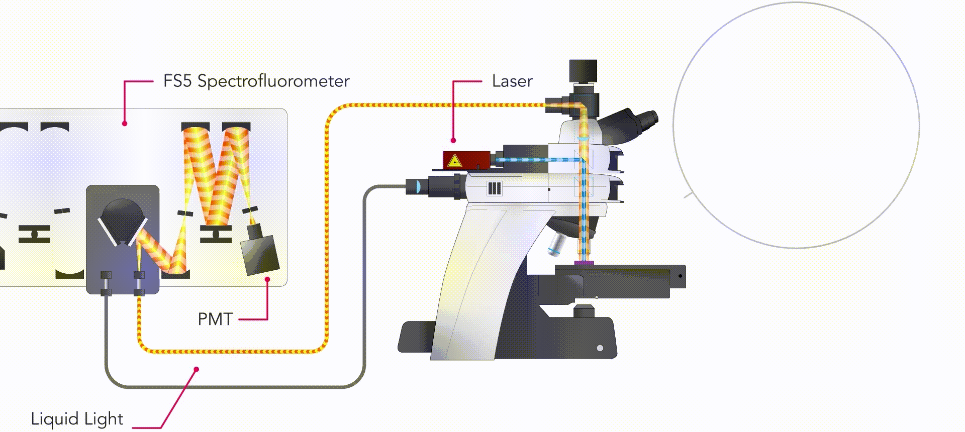
Figure 5: MicroPL configuration for lifetime mapping.
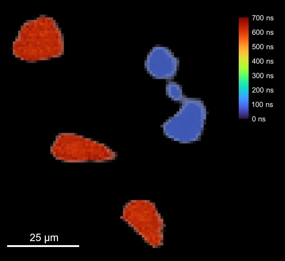
Figure 6: Photoluminescence lifetime map of the phosphor microparticles.
