Confocal Raman microscopy combines the spectral information from Raman spectroscopy with the spatial filtering of a confocal optical microscope for high-resolution chemical imaging of samples. The spectral Raman information is sensitive to the vibrational modes of the sample and provides extensive chemical, physical and structural insight, while the confocal optics of the microscope enable the analysis volume within the sample to be spatially filtered with high resolution in both the lateral (XY) and axial (Z) axes. The synergy between the spectral and spatial information enables the chemical analysis of individual particles, discrete sample features or layers down to less than 1 μm in size.
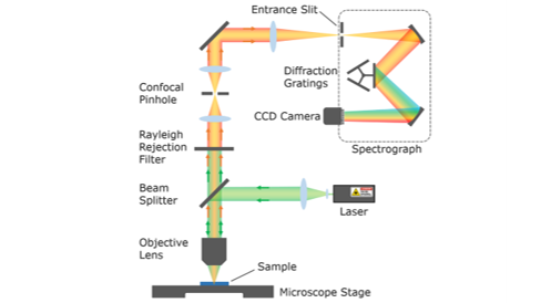
A schematic of the optical layout of a confocal Raman microscope is shown in Figure 1. The sample to be analysed is placed on the confocal microscope stage and the excitation light from a laser is reflected by the beam splitter and focused down onto the sample through the objective lens of the microscope. The Rayleigh and Raman scatter from the sample are collected by the objective lens, transmitted through the beam splitter and passed through the rejection filter which removes the Rayleigh scatter. The remaining Raman scatter is then focused through a confocal pinhole and enters the spectrograph where it is wavelength separated and the spectrum detected using an array detector. Spatial information is obtained by moving the microscope stage, and therefore the sample, in the XYZ directions and acquiring an array of Raman spectra which are then used to generate three dimensional Raman spectral maps of the sample.

Figure 1: Optical Layout of a Confocal Raman Microscope.
Lasers are the ideal excitation source for Raman microscopy as they provide high intensity light which increases Raman scattering and sensitivity, are monochromatic which gives high spectral resolution, and have low divergence; enabling them to be focused to a small spot on the sample for high spatial resolution. The wavelength of the laser is important as it influences the Raman scattering intensity, spatial resolution and background fluorescence. For this reason, high-end Raman microscopes, such as the Edinburgh Instruments RM5, can hold multiple lasers simultaneously so that the most appropriate laser wavelength for each sample can be quickly switched to using the software. For a more detailed discussion of the effects of laser wavelength read our post on how to choose laser wavelengths.
The objective lens is used to both focus the laser excitation onto the sample and collect the Raman scatter from the sample. The choice of objective lens is important as the numerical aperture (NA) of the objective lens and the laser wavelength together determine laser spot size on the sample and the spatial resolution of the Raman mapping. The theoretical lateral resolution that can be achieved is diffraction-limited and given by the Rayleigh criterion:

Increasing the NA (increased magnification) of the objective lens improves the spatial resolution. For example, a 10x objective with an NA of 0.25 and 532 nm excitation light would have a lateral resolution of ~1300 nm while a higher magnification 100x objective with an NA of 0.90 could in theory achieve a lateral resolution down to ~360 nm.
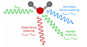
Only around 1 in 10 million of the scattered photons are Raman scattered with the rest being Rayleigh scattered. The intense Rayleigh scatter contains no useful sample information and must be discarded prior to detection which is achieved by passing the scattered light through either an edge or notch rejection filter.
Edge filters are sharp long pass optical filters which absorb all wavelengths up to their ‘edge’ and then transmit all wavelengths beyond that edge. The edge wavelength is chosen so that the Rayleigh scattered light is strongly absorbed and the Stokes Raman scattered light is transmitted. They enable lower Raman shift wavenumbers to be observed compared to notch filters but only allow the Stokes lines to be observed.
Notch filters are holographic filters which have a sharp absorption peak at one particular wavelength, chosen to coincide with the laser wavelength, and transmit all other wavelengths. The advantage of notch filters is that the Stokes and anti-Stokes Raman scatter can be observed simultaneously. The downside is that they have a broader absorption profile preventing low wavenumber peaks from being observed and have a finite lifetime under laser irradiation and therefore must be replaced periodically.
The confocal pinhole is the defining feature of a confocal microscope and is used to improve the spatial resolution, increase the contrast, and decrease the fluorescence background of the Raman mapping. The confocal pinhole blocks out of focus Raman scatter from entering the spectrograph and being detected, as illustrated in Figure 2. Without a confocal pinhole (left) both the in-plane Raman scatter (red) and out of plane Raman scatter (blue) enter the spectrograph and would be detected. In contrast, the confocal pinhole (right) blocks the out of plane Raman scatter from entering the spectrograph.

Figure 2: Role of the confocal pinhole in blocking out of focus Raman scatter.
The confocal pinhole functions as a spatial filter in the axial (Z) direction by blocking the Raman scatter from above and below the focal plane (extrafocal) and is essential for 3D mapping. Without the pinhole, there is no axial spatial filtering and Raman scatter from the entire axial volume of the sample would be collected with no axial resolution. The pinhole is also beneficial for lateral 2D mapping since it enhances the contrast by blocking extrafocal scatter and background fluorescence and also marginally improves the lateral (XY) resolution.
The terms truly confocal and pseudo confocal are frequently encountered when reading about Raman microscopes. Truly confocal microscopes, such as the RM5, have a physical pinhole in the confocal plane of the microscope and offer superior confocal performance, while pseudo confocal microscopes simulate the effect of a pinhole using the orthogonal arrangement of the entrance slit of the spectrograph and the pixels of the CCD camera.
The spectrograph uses a combination of mirrors and a diffraction grating to spatially separate the different wavelengths of the Raman scatter and image them onto an array detector for detection. The focal length of the spectrograph, the width of the entrance slit and the groove density of the diffraction grating determine the spectral resolution of the Raman microscope which dictates how effective the microscope is at resolving closely spaced Raman peaks. The groove density of the diffraction grating has the largest influence on the spectral resolution with spectral resolution increasing with increasing groove density. However, there is a trade-off as the higher the groove density, the narrower the spectral range that the grating is effective over. Spectrographs therefore typically contain multiple gratings, with a range of groove densities, installed on a rotating turret. The low groove density gratings can then be used to acquire the entire spectrum out to high wavenumbers and the high groove density grating used for spectrally resolving closely spaced peaks over a narrower region of the spectrum.
The Raman scatter is detected using an array detector which captures the spectrum in a single acquisition. The most common array detector is a CCD camera which is a high sensitivity camera with a rectangular array of pixels that the Raman spectrum is imaged onto and converted into an electronic signal. An EMCCD (Electron Multiplying CCD) is similar to a regular CCD but contains an additional on-chip electron-multiplying amplification stage that enables faster data readouts while preserving the high sensitivity and are the preferred detector for rapid Raman mapping with short acquisition times. One limitation of CCD cameras is that they are only sensitive out to ~1000 nm making them unsuitable for Raman microscopy with 1064 nm excitation. For 1064 nm excitation an InGaAs linear array detector which has maximum sensitivity in the near-infrared region is used in place of the CCD.
You can find out more about our RM5 Raman Microscope on our website. If you have enjoyed reading this article and wish to learn more about our confocal microscope, our applications and products then why not contact a member of our team at sales@edinst.com or follow us on social media via your preferred channel below.