


From Maps to Memories: Two Years in Review
Map of the Month is a fantastic way for us to highlight features in our instruments and software in a more light-hearted way. We love to see the scientific images we can create using a range of techniques such as Raman, fluorescence lifetime, photoluminescence, second harmonic generation and more.
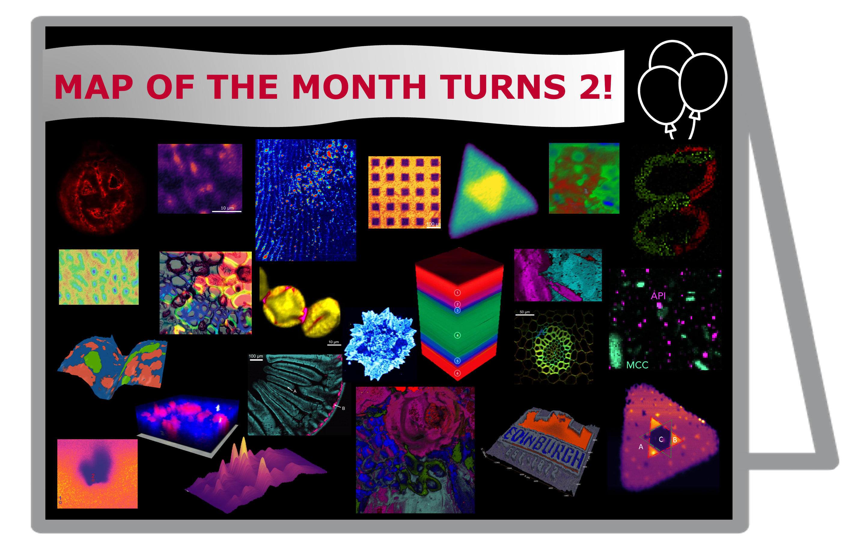
This month, to celebrate the blogs 2nd birthday we asked our team at EI to reflect on the past couple of years and pick their favourite map so far…
Alix Bailie – Applications Scientist
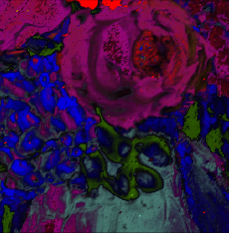
 My favourite map comes from February this year, which looked at the distinctive pigments in historical art pieces. It’s the most interesting intersection of art and science! You can see from the white light and false colour Raman images how the piece came together in a totally different light. Not only does it tell you what the paint these artists used could have been made from, but it helps historians in restoration processes which makes all these works available to enjoy for the future.
If you’ve ever visited a museum while they’ve been restoring a piece several meters in size, you’ll see how important it is that areas requiring care match the original work as close as possible.
CJ Seller – Head of Marketing
 I have chosen the most recent one – Document Forgery. I am new to Edinburgh Instruments, starting in the summer as the Head of Marketing. The Map of the Month is an engaging piece that shows our products’ wide-ranging features and applications. The blog is engaging to read and was a part of my research when I joined the company! My personal favourite (so far) is this month’s which featured the use of Raman PL mapping for tracing document forgery.
I had no idea that the forensic analysis included potentially using Raman mapping – and that it could be a relatively simple solution to unveil the lengths criminals go to when hiding their crimes. As an avid fan of true crime, it led me down many other paths… how else can this application and product help solve crimes?? The image of the two eights – one to the naked eye, and the other under the Raman microscope was striking and would certainly make convincing evidence in a court of law. I find that as a non-scientist, seeing the products and applications applied to everyday life is a brilliant and fascinating way to show applied science. 
Grant Cumming – Applications Scientist
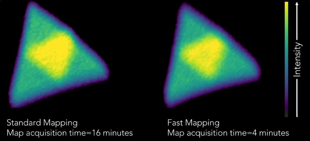
 I have chosen October 2022 – Fast Mapping of MoS2. As a scientist who has worked for years acquiring high-resolution Raman maps – while lacking Edinburgh Instrument’s Fast Mapping feature – I have spent many late nights in the lab. The ability to reduce the total map acquisition time by up to 75% would have been amazing. It’s the ability to increase sample size or achieve reasonable home times represented by this map which led me to choose it as my favourite; albeit with a retrospective jealousy.
While the molybdenum disulfide sample may not be as visually complex as some of our other Maps of the Month, it still produces a striking map with all of the same chemical information whether imaged without Fast Mapping in 16 minutes, or with Fast Mapping in 4 minutes.
Paula Duffy – Marketing Executive & Alistair Rennie – Group Head of Product
Paula and Alistair both chose December 2021 as their favourite Map if the Month, featuring a Fluorescence Lifetime image (FLIM) of a stained pine tree section.
 What makes this map so memorable to you Alistair? At the time we had recently launched our new Raman line of products and I remember this image being one of the first that truly impressed me. It was so eye catching and thought provoking, but more than that, made me realise how powerful the multimodal RMS1000 Raman Microscope really is.
 What it is about the sample that caught your attention, Paula? The map itself is really interesting to look at because of the colours and shapes – you wouldn’t believe this is what a pine needle actually looks like! I think this is a fantastic example of how a sample can look so different beyond what meets the eye. Using various imaging techniques to reveal the details of structures at the cellular level gives us a real appreciation for the beauty of nature and how technology can help us understand how plant cells function.
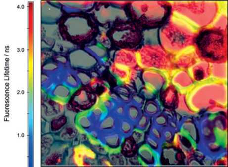
Alistair, why was the FLIM feature so exciting for you? It brings together two expertise of EI into one instrument, Raman and Fluorescence. Moreover, I think kudos to our software team for writing the code to make this type of analysis possible in one programme.
Contact Us