
Laser Excitation Diversity
Up to three fully software controlled internal lasers from 405 nm – 1064 nm, minimising footprint, maximising versatility


Ramacle® is an all-encompassing software package written for ease of use and complete operation of the RM5.
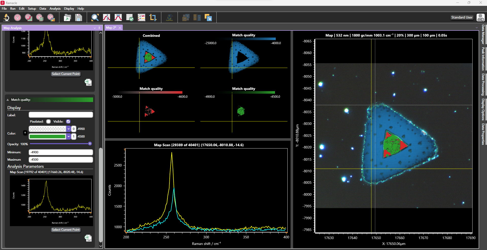
The software prioritises automation, reducing the need for manual adjustments and allowing users to focus more on sample analysis. From initial microscope setup, where users visualise their sample in a spacious window, to configuring laser parameters, pinhole settings, and grating selection for spectral acquisition, Ramacle handles all aspects seamlessly.
On the RM5, Ramacle comes with standard data acquisition methods such as single measurements, multiple and accumulated scans, kinetic scan, as well as a comprehensive suite of mapping techniques when upgraded to a motorised stage e.g. 2D, 3D, SurfMAP, and FastMAP. Upgrades and accessories such as temperature stages and multiwell plates unlock additional software features.
Intuitive use is at the core of Ramacle’s design. We begin by focusing your sample using the microscope setup. Once you’re satisfied with the microscope image, we can proceed to the measurement setup window. The white-light image will carry over, allowing you to click on different areas of the sample for live Raman feedback using the live option. This feature helps you verify your measurement parameters and focus. You can then take single or multiple spectra from the selected sample area.
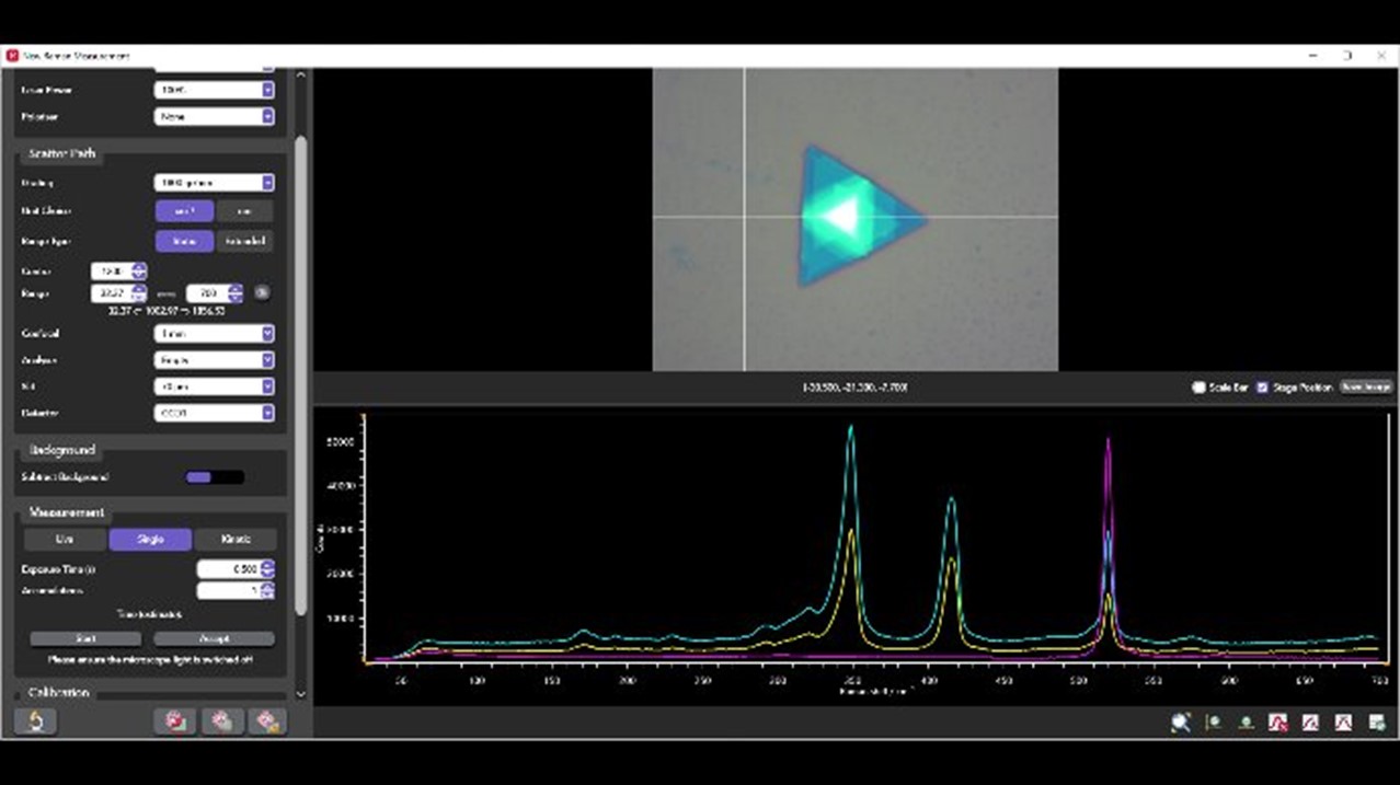
Figure 1: Ramacle Software
Additionally, kinetic series can be set up simply in Ramacle, and if using a temperature stage, temperature ramps and spectral series can also be set. The RM5 can be configured to acquire photoluminescence emission spectra. Ramacle measures spectral PL responses using the CCD camera enabling single point and mapping data to be collected. Measurements are made by selecting a low groove density grating to cover the entire emission range. The software can be operated in both wavenumber and wavelength scale making working in Raman and PL effortless.
2D mapping in Ramacle allows you to track the distribution of components in your sample. All the user needs to do is determine the area of interest to be investigated. Using Ramacle the area of interest to be mapped is defined, including areas larger than the field of view using stitched images. On the microscope image the map dimensions and step size are set for XY measurements, which will then move the stage to take a Raman spectrum from each point.
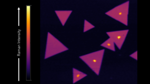
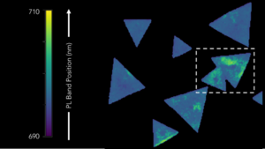
Figure 1: Raman and photoluminescence imaging of MoS2
Raman and PL imaging can be carried out on the same sample to provide complementary information, such as in 2D materials. In the example shown MoS2 is first imaged to study the Raman spectra, which indicates layer number. Then by simply changing the grating the PL emission is revealed, which can be used to measure strain across the sample.
Ramacle operates 3D mapping similarly to 2D mapping, with the added ability to define the map along the Z-axis. Thanks to the truly confocal nature of the RM5, highly resolved 3D maps can be captured. This advanced capability extends Raman mapping beyond surface analysis, constructing a detailed 3D chemical image at the micron scale.
The Ramacle software can then display the result focusing on one 2D layer, on the 2D stacks collected, or as a full volumetric. Shown below are two common examples for 3D mapping; first 3 µm polystyrene beads, and on the right we a see an inclusion in pegmatite. Green is quartz, red is anatase, and blue is an unknown inclusion.
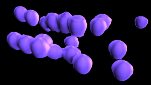
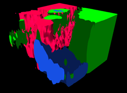
Figure 1: 3D Mapping of polystyrene bead and pegmatite
The SurfMAP® feature of Ramacle enables the user to analyse difficult samples with rough and uneven surfaces. This is done by adjusting the Z-axis to ensure the laser remains in perfect focus across the sample surface. Without this feature non-flat surfaces cannot be accurately mapped due to variation in signal caused by the laser moving out of focus to the sample.
For example, when a pharmaceutical tablet has an indentation, a lab using Raman or PL imaging to inspect the tablet’s surface after manufacturing must account for the uneven surface.
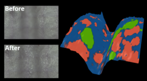
Figure 1: Raman surface mapping of a pharmaceutical tablet.
The resulting map can be displayed in 3D showing the sample height variation and, in this example, reveals the distribution of paracetamol (blue), aspirin (coral), and caffeine (green) in the tablet.
The RM5 is built with flexibility in mind. A choice of excitation lasers and associated laser rejection filters (both edge and notch) are available depending on application requirements.

Gratings are chosen for optimum resolution for each laser excitation, with up to a maximum of five gratings per system.

A choice of CCD, EMCCD and InGaAs detectors are also available dependent on requirements, with a maximum of two detectors being integrated per system.
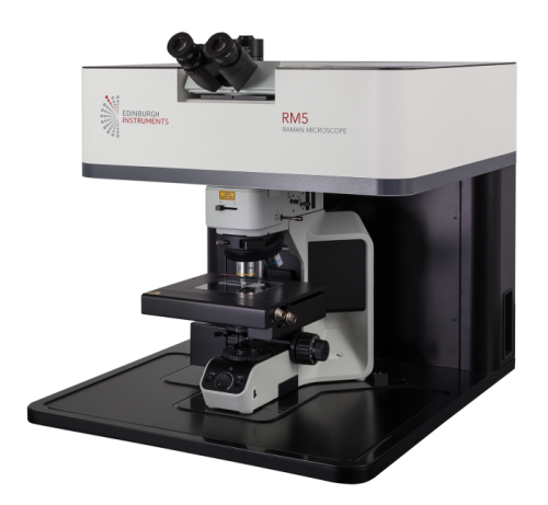
The RM5 uses one of the most modern microscopes on the market for first class Raman microscopy. You can use the microscope beyond pure Raman microscopy; the RM5 has been designed to maintain the full capability of the microscope allowing all the necessary tools to be added for exceptional visualisation and contrast of your samples.
Brightfield, darkfield, polarised light, differential interference contrast (DIC) and fluorescence are all available. Alongside a choice of high quality microscope objectives, a high performance camera can be added to the microscope to ensure pictures of your samples (and associated Raman maps) are captured with excellent quality and resolution.

Other accessories such as a polarisation kit and a Class I laser safety enclosure are also available to further expand the capabilities, flexibility and safety of your RM5 system.
