


Pesticide Detection on Apple Skin Using SERS
Introduction
Protecting food crops is vital to the world’s food chain, and pesticides are a critical tool to kill, repel, and control pests. Two million tonnes of pesticides are used annually worldwide, with this value predicted to increase over time.1 However, pesticides can cause severe environmental issues to aquatic systems, wildlife (e.g., bees), air, and soil. Additionally, the reported effects of pesticides on human health range from short-term, such as skin irritation and headaches, to chronic effects, such as asthma and cancer. There are also concerns about the consequences of consuming pesticides from the small, but repetitive, doses in our diet. 2–4
One method to reduce the dangers of pesticides is limiting the allowed concentration used on crops. This is controlled by analysing the crop and determining the level of pesticide present. Apples are one of the most pesticide-treated fruits and as such producers have a list of regulations they must abide by for sale to the public.
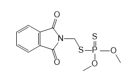
Figure 1: Chemical structure of phosmet.
Phosmet is an organophosphate insecticide used on apple trees to control codling moths, the 2018 Code of Federal Regulations set the pesticide residue tolerance on apples of phosmet as 10 ppm. High-performance liquid chromatography, mass spectrometry, and gas chromatography are currently the most used techniques for testing down to low concentrations. However, these are time-consuming and costly, and Raman spectroscopy offers several advantages such as rapid and non-destructive fingerprint-like identification with little to no sample preparation.
Surface enhanced Raman scattering (SERS) is an enhancement technique used with Raman spectroscopy to provide lower limits of detection. SERS offers a signal enhancement of up to 10^10-10^15 and also advantageously quenches the fluorescence of analytes. Commercial SERS substrates are readily available, making SERS an accessible technique for low-concentration detection. In this application note, residual phosmet insecticide on apple skin is detected using SERS.
Materials and methods
An RM5 Raman Microscope equipped with a 785 nm laser and a 600 gr/mm grating was used to analyse the phosmet samples. Gold SERS substrates from Hamamatsu were used to provide the enhancement effect. Since gold nanoparticles typically absorb in the region of 520 nm – 580 nm the preferred excitation wavelength for gold SERS substrates is standardly 785 nm.
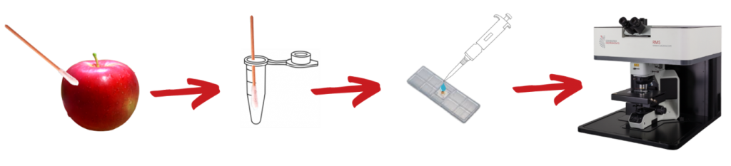
Figure 2: Experimental workflow for apple skin analysis.
Aqueous solutions of 10 ppm and 1 ppm phosmet were prepared, and 6 µL of each phosmet solution was pipetted into the well of the SERS substrate, left to dry, and analysed. To create a contaminated apple sample, a small volume of 10 ppm phosmet solution was dropped onto a cleaned, cut-out section of apple skin and left to dry. Once dry a wet cotton bud was used to swab the surface of the apple skin and placed into a small volume of water. 6 µL of this sample was pipetted onto the SERS substrate for analysis.
The SERS substrates were analysed by placing underneath the Raman microscope and exciting the sample with a 785 nm laser using an exposure time of 5 seconds. As no spatial resolution was required, the pinhole diameter was set at 2 mm to maximise the Raman signal.
Results and Discussion
Figure 3 shows the Raman spectra of the 4 samples analysed. The 10 ppm solution was analysed in a cuvette meaning there was no SERS enhancement; this measurement was performed to show the standard Raman response from phosmet at a low concentration. Without enhancement from the SERS substrate, the 10 ppm solution of phosmet has very little Raman signal, however, when deposited on the gold SERS substrate a strong Raman spectrum is observed. When further diluted to 1 ppm a pronounced Raman signal is still acquired revealing the low detection limit achievable with SERS.
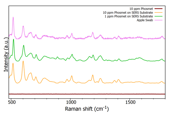
Figure 3: Raman spectra from three phosmet solutions and a spiked apple skin swab.
The spectrum shown in pink in Figure 3 is the swabbed apple skin spiked with 10 ppm phosmet. The spectrum is noisier than the 10 ppm solution of phosmet however well-defined peaks are still observed, peak assignments are detailed in Figure 4.
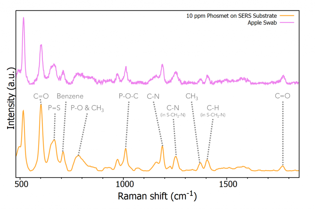
Figure 4: Raman spectrum from 10 ppm phosmet solution and spiked apple skin swab.
Band assignments in Figure 4 highlight how sensitive the fingerprint region of a Raman spectrum is to specific functional groups allowing pesticide identification.5 Most of the peaks observed in the 10 ppm solution spectrum are also easily visible in the apple swab spectrum. Whilst the peaks are generally less intense and noisier, the Raman spectrum is easily identifiable as phosmet after smoothing and normalisation.
Conclusion
This study demonstrated a non-destructive sampling and analysis technique for monitoring low concentrations of pesticides on apple skin. Gold SERS substrates were used to enhance the Raman spectrum of a phosmet, proving effective at identifying concentrations at the residual tolerance level set by the Code of Federal Regulations.
References
- Sharma, A. et al. Worldwide pesticide usage and its impacts on ecosystem. SN Appl. Sci. 1, (2019)
- Kim, K.-H., Kabir, E. & Jahan, S. A. Exposure to pesticides and the associated human health effects. Sci Total Environ. (2017)
- Bonner, M. R. & Alavanja, M. C. R. Pesticides, human health, and food security. Food Energy Secur. 6, 89–93 (2017)
- Rani, L. et al. An extensive review on the consequences of chemical pesticides on human health and environment. Clean. Prod. 283, 124657 (2021)
- Jiang, L. et al. Rapid detection of pesticide residues in fruits by surface-enhanced Raman scattering based on modified QuEChERS pretreatment method with portable Raman instrument. SN Applied Sciences. (2019)
Keep in Touch
If you are working in the field of pesticide residue detection and have enjoy this application note, be sure to subscribe to our eNewsletter or follow us on social media using the links below.
