


Graphene Characterisation Using Raman Microscopy
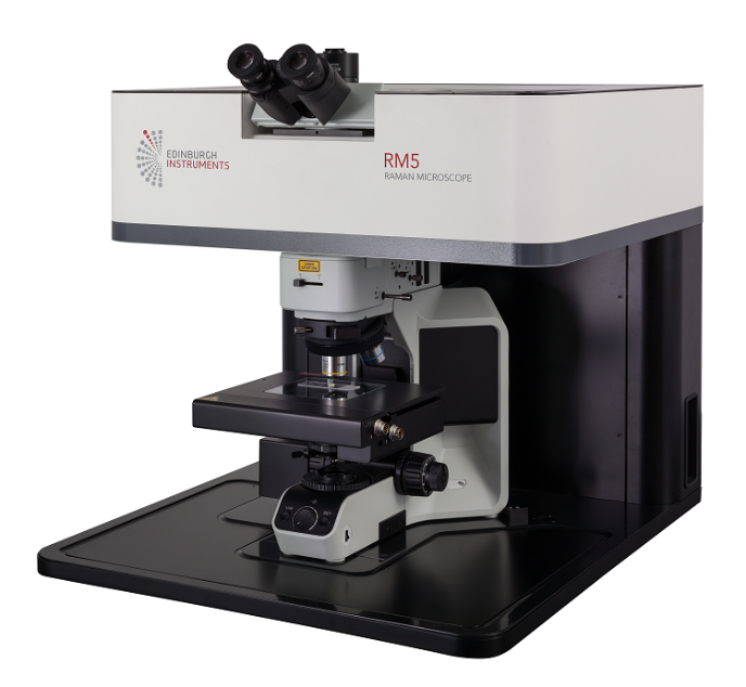
In this virtual demo, we show how to perform high-resolution mapping and analysis of graphene films using the RM5 Raman Microscope and Ramacle software.
In it, we show how to identify and interpret:
Layer number (monolayer, bilayer, etc.)
Defects and disorder (D band intensity and distribution)
Strain and stress patterns (G and 2D band shifts)
This tutorial covers the full workflow, from sample setup and spectral acquisition to advanced data analysis and visualisation. It's ideal for materials scientists, nanotechnology researchers, and Raman spectroscopy users aiming to extract more from their graphene data.
Don’t forget to like, share, and subscribe for more Raman spectroscopy tutorials and materials analysis content.
Raman & FLIM Imaging Tutorial: RaFLIM® using the RMS1000 Confocal Microscope Guide
Do you need help performing Raman and FLIM images of the same part of your sample? This step-by-step tutorial covers everything you need to know.
• FLIM setup: learn how to set laser and detector conditions in Ramacle® for fluorescence and phosphorescence lifetime, and then perform imaging with those conditions.
• FLIM analysis: create images based on fluorescence intensity or lifetime, with up to four exponential component fits in Ramacle®. • Raman setup: learn how to set laser and scatter path conditions in Ramacle® for Raman imaging.
• Raman analysis: create images based on various Raman spectral conditions on the same sample area or volume that you performed FLIM on!
Thanks for watching, remember to like, comment and subscribe for content like this and more!
Chapters:
0:00 – Intro
0:35 - Focusing our Sample
1:34 - PL Spectra Measurements
2:22 - Selecting Optimum Wavelength for FLIM Measurements
2:49 - Lifetime Measurements
3:38 - Lifetime Measurement Analysis
4:00 - Performing 2D Mapping - FLIM
6:50 - Performing 2D Mapping - Raman
8:12 - Outro
Visit our Website: www.edinst.com
Raman and beyond: RaFLIM® Feature Highlight
Discover the possibilities beyond Raman measurements with the RaFLIM® using the RMS1000. Building on over 50 years of fluorescence expertise, Edinburgh Instruments, FLIM upgrade integrates seamlessly with Raman capabilities. Powered by our intuitive all-in-one software package, Ramacle, you can effortlessly acquire and analyse RaFLIM® data.
To stay up to date with all things Raman and Fluorescence be sure to like and subscribe. Leave a comment down below if you have any questions or what you would be interested in seeing next.
How to Setup and Use a Temperature Stage with Edinburgh Instruments RM5 and RMS1000 Raman Systems
Do you need help setting up your Linkam temperature stage for Raman microscopy?
This step-by-step tutorial covers everything you need to know—from installation to automating temperature control and spectral acquisition in Ramacle® - using the RM5 or RMS1000 Raman Microscope.
What’s in this video?
Installation: Learn how to properly mount the THMS600 stage and connect the necessary cables for precise temperature control.
Software Setup: Discover how to configure temperature controlled measurements in Ramacle®.
Whether you're working with high or low-temperature experiments, this guide will help you maximise your setup’s potential for temperature dependent Raman research.
Thanks for watching, remember to like, comment and subscribe for content like this and more!
#LinkamTHMS600 #RamanMicroscopy #Tutorial #Science #Laboratory #MyEdinburghInstruments #TeamRaman
0:55 Introduction
0:55 Remove Transit Screws
01:19 Controller Connections
02:10 Attaching the Mounting Plate
02:50 Mount on Microscope Stage
03:10 Fill Liquid Nitrogen Dewar
03:38 Feedthrough Cables
03:58 Stage Connections
04:45 Loading a Sample
05:19 Connect to Ramacle
05:40 Purging the Stage
06:46 Sample Focusing
07:15 Temperature Scan
09:29 Temperature Trend Analysis
09:50 Summary
Enhance Your Raman Analysis: Database Searching in Ramacle with KnowItAll
In this video, we explore the integration of KnowItAll with our Ramacle® software, enhancing your Raman analysis. Learn how the KnowItAll search function is seamlessly incorporated into our RM5 and RMS1000 Raman systems. This integration speeds up material identification, provides access to a comprehensive material library, and improves the accuracy and efficiency of your data analysis.
Searching the database is quick and easy, making your workflow more efficient.
0:00 Introduction
0:26 Benefits of KnowItAll Software
0:49 Taking a Spectral Measurement for Analysis
0:57 Ramacle - Material Assignment Window
1:30 Setting up Raman Database - within KnowItAll
2:03 Selecting Materials for Comparison
2:17 Importing Selected Material to Ramacle
2:29 Viewing data for future Reference
2:35 Outro
Whether you're a scientist, researcher, or industry professional, this upgrade is designed to take your analytical work to the next level.
To see more content like this and more consider subscribing to stay up to date.
Polymer identification made easy: Using ATR-FTIR and KnowItAll Material Library
In this demo we show how ATR-FTIR can identify a polymer using our Miracle software with the KnowItAll upgrade on an IR5 FTIR Spectrometer.
0:00 Introduction
0:35 Using the IR5 with Miracle
0:51 Taking a Background Measurement
1:12 Setting up the ATR Attachment for the IR5
1:45 Analysing an Unknown Polymer Sample
1:59 How to Use KnowItAll Software
3:26 Reading Measurements Storage
3:30 Outro
If you enjoyed the video, please give it a like, and don't forget to subscribe for more insights on material science and analysis. Got a question or a topic you'd like to see next? Drop a comment below! And make sure to click the bell icon to stay updated on our latest videos.
What is Raman Spectroscopy?
Welcome to our Introduction to Raman Spectroscopy!
In this video, we delve into the fascinating world of Raman Spectroscopy, an essential analytical technique used in various fields such as chemistry, physics, and materials science. This video will introduce you to the fundamental principles and applications of Raman spectroscopy.
0:00 Intro
0:13 Looking at a Sample
0:21 C.V Raman Discovery
0:41 Stokes and Anti-Stokes
1:00 Jablonski Diagrams
1:33 Unique Molecular Vibrations
1:45 Measuring the Raman Spectrum
2:01 Summary of Raman Spectroscopy
2:24 Outro
Be sure to subscribe to stay up to day with more videos like this from Edinburgh Instruments. We hope you enjoyed.
How to Take The Perfect Raman Spectrum
In this tutorial video, we guide you through the process of acquiring an excellent Raman spectrum on the RM5 confocal microscope. We cover what parameters make a great spectrum and how each component within a Raman microscope can be used to enhance the quality of your spectrum based on your application needs.
Whether you are a beginner in the field or an expert looking to refine your skills, this video offers insights and practical tips to ensure that you can always get the most out of your spectral measurements.
0:00 - 0:30 Introduction
0:30 - 0:47 What makes a good spectrum?
0:47 - 1:06 Spectral resolution
1:06 - 1:28 Spectral sensitivity
1:28 - 1:50 Spectral range
1:50 - 2:09 The need for balance
2:09 - 2:31 Fluorescence
2:31 - 3:11 How to control spectral quality
3:11 - 5:46 The laser
5:46 - 6:41 The diffraction grating
6:41 - 7:18 The confocal pinhole
7:18 - 7:39 The slit
7:39 - 8:57 Summary
Speaking Spectroscopy Ep 1: ICAVS
In this episode of Speaking Spectroscopy Angela and Matthew reflect on ICAVS.
Hear what they have to say about the latest development and research in Raman.
RM5 Raman Mapping Virtual Demo
Raman mapping is a powerful technique that allows chemical and structural information to be imaged on a sample.
This Virtual Demonstration shows how easy it is to perform a mapping experiment on the RM5.
Each step between switching on the system and producing a processed Raman map is explained.
0:00 Intro
0:14 Setting up the RM5
0:45 Calibrating the Microscope Stage
1:07 Loading of the Sample
1:30 Focusing on The Sample
2:00 Performing Spectroscopic Analysis
3:00 Setting up and Running Mapping
4:48 Map Analysis
5:51 Summary
This or That: Raman and IR Edition
This or That - Raman vs IR edition.
IR and Raman are complimentary vibrational spectroscopy techniques. So how do we pick which one to use? A lot of the time your samples make that decision, and our latest blog explains in more detail why!
https://www.edinst.com/blog/infrared-or-raman-spectroscopy/
#Raman #FTIR #ScienceShorts #Shorts
Raman Microscopy: A Versatile Analytical Tool for Art Authentication and Forensic Analysis
Raman Microscopy can be used for the identification and analysis of forensic and cultural materials. In this poster presentation, discover how the RMS1000 and RM5 Raman microscopes can be used for forensic investigations such as drug detection, bank note authenticity and archaeological analysis.
2D Materials Multimodal Characterisation in 60 seconds #shorts
Watch the full video for all the details: https://www.youtube.com/watch?v=BZ9rDfdpwTU
Looking to fully characterise your 2D Materials using one instrument?
Our RMS1000 Raman Microscope enables Multimodal Imaging, allowing complete characterisation of your materials.
Watch our full video to discover how the RMS1000 can be used for:
#Raman
#Photoluminescence
#SecondHarmonicGeneration
and more: https://www.youtube.com/watch?v=BZ9rDfdpwTU&t=3s
#shorts #Shorts
2D Materials Multimodal Characterisation with Raman Microscope
Interested in fully characterising your 2D materials with one instrument?
Watch our poster presentation that will show you how Raman, Photoluminescence, and Second Harmonic Generation Imaging can be employed on the RMS1000 the determine layer thickness and stacking of a tungsten diselenide crystal.
#Raman #2DMaterials #graphene #multimodal
Raman Mapping with Edinburgh Instruments
🗺️ Raman Map your sample with Edinburgh Instruments Raman Microscopes.
💻 Ramacle – One powerful intuitive software for Raman Mapping with confidence.
🚀 Raman and Beyond – The RMS1000 can drive your research beyond Raman thanks to its upgradeable open architecture design.
🔬 Benchtop Raman – The RM5 suits every Raman application with ease-of-use at its core.
Products:
RM5 Raman Microscope: https://www.edinst.com/products/rm5-raman-microscope/
RMS1000 Raman Microscope: https://www.edinst.com/products/rms1000-raman-microscope/
#microscopy #raman #microscope
Raman Microscopy Poster Presentation Series – Food & Drinks Industry
In this short presentation we will present our poster on the use of Raman spectroscopy for food analysis. We will discuss how our RM5 and RMS1000 Raman Microscopes can help regulatory bodies with food and beverage analysis.
Raman Microscopy RAMACLE® Software Highlights (Various)
Edinburgh Instruments has been providing high performance instrumentation in the Molecular Spectroscopy market for almost 50 years. Our Precision Raman microscopes continue our commitment to offering the highest quality and sensitivity instruments.
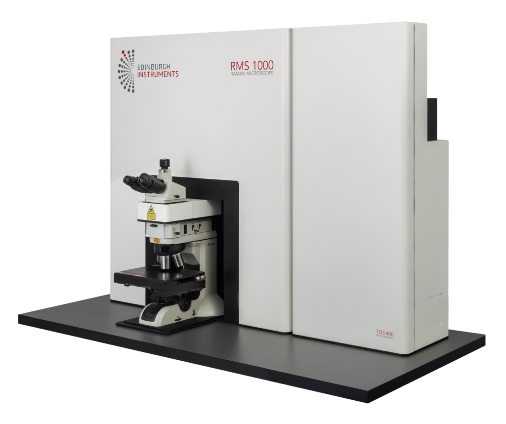
The RMS1000 is an open architecture research grade confocal Raman microscope. It has been designed so it can be adapted to almost any modern, state-of-the-art Raman application. This high-end research tool has been built with no compromises; resulting in a system that stands alone in both specification and ease of use. Applications beyond Raman such as time-resolved fluorescence microscopy and fluorescence lifetime imaging (FLIM) are all possible with the versatile RMS1000.
The RM5 is a compact and fully automated Raman microscope for analytical and research purposes. The truly confocal design of the RM5 is unique to the market and offers uncompromised spectral resolution, spatial resolution, and sensitivity.
In this video we take a look at the capabilities provided by the RMS1000 and RM5 in conjunction with RAMACLE® software.
2D Mapping
(RMS1000, RM5 & Ramacle Software):
Painting
PL Mapping of Perovskite
Fast Mapping
(RMS1000, RM5 & Ramacle Software):
MoS2
Surface Mapping
(RMS1000, RM5 & Ramacle Software):
Carbon Fibres
Medicinal Tablet
3D Mapping
(RMS1000, RM5 & Ramacle Software):
Transdermal Patch
FLIM
(RMS1000, RM5 & Ramacle Software):
Mouse Intestine
Multimodal Imaging
(RMS1000 & Ramacle Software):
WSe2
Visit our website for further information on our Raman Microscopes: https://www.edinst.com/types/raman-microscopes/
Ramacle® Software Highlight: Fast Mapping
This Software Highlight focuses on Fast Mapping in our Ramacle® software.
Fast Mapping enables the user to acquire Raman maps with significantly reduced acquisition times via more efficient stage movement. Raman Fast Mapping can take significantly less time than standard mapping, without compromising the quality of the map.
More about Ramacle®: The software provides control, visualisation, data acquisition, analysis and presentation of Raman microscopes whether it is used for generating Raman spectra or with advanced upgrades such as Raman mapping.
Ramacle® enables sample visualisation, live signal monitoring and parameter optimisation before every measurement. The instrument status and signal are displayed and constantly updated during measurements.
Data generated by Ramacle® have a proprietary file format. This contains all measurement and instrumental properties, allowing the user to retrieve important information whenever needed
and ensures data traceability. Simple input and output functions provide the required compatibility with third party data analysis or presentation packages.
KnowItAll™ Raman Identification Pro spectral library is available for material identification and advanced analysis. Data acquisition methods such as single measurements, multiple and accumulated scans, kinetic scans and generation of maps (accessory dependent) are implemented by intuitive and in user-friendly wizards.
Find out more about Ramacle® on our website: https://www.edinst.com/us/products/ramacle-software/
Surface Mapping Software Highlight: https://www.youtube.com/watch?v=OAQcSXVt1Fc&t=5s
0:00 Intro
0:14 Raman Mapping
0:40 Standard Mapping
1:46 Fast Mapping
2:16 Mapping Comparison
Raman Spectroscopy - Capabilities Beyond Raman
Providing world-class Molecular Spectroscopy solutions for over 50 years, Edinburgh Instruments precision Raman microscopes offer the highest quality and sensitivity instruments.
RMS1000 Research Grade Microscope:
Extending capabilities beyond Raman, the RMS1000 confocal Raman Microscope stands alone in both specification and ease of use. Applications beyond Raman such as time-resolved fluorescence microscopy and fluorescence lifetime imaging (FLIM) are all possible with the versatile RMS1000. An open architecture research grade microscope, it can be adapted to almost any modern, state-of-the-art Raman application.
RM5 Analytical & Research Grade Raman Microscope:
A fully automated and fully integrated Raman microscope for analytical and research purposes, the RM5 with its truly confocal design is unique to the market. It offers uncompromised spectral resolution, spatial resolution and sensitivity.
Discover our range of Raman Microscopes online: https://www.edinst.com/types/raman-microscopes/
Ramacle® Software Highlight: Surface Mapping
This Ramacle® Software Highlight focuses on Surface Mapping detailing why it is a necessary feature and how it works. Surface Mapping allows the user to analyse tricky, uneven sample types.
The software provides control, visualisation, data acquisition, analysis and presentation of Raman microscopes whether it is used for generating Raman spectra or with advanced upgrades such as Raman mapping.
Ramacle enables sample visualisation, live signal monitoring and parameter optimisation before every measurement. The instrument status and signal are displayed and constantly updated during measurements.
Data generated by Ramacle have a proprietary file format. This contains all measurement and instrumental properties, allowing the user to retrieve important information whenever needed
and ensures data traceability. Simple input and output functions provide the required compatibility with third party data analysis or presentation packages.
KnowItAll™ Raman Identification Pro spectral library is available for material identification and advanced analysis. Data acquisition methods such as single measurements, multiple and accumulated scans, kinetic scans and generation of maps (accessory dependent) are implemented by intuitive and in user-friendly wizards.
Find out more about the software: https://www.edinst.com/products/ramacle-software/
0:00 Intro
0:09 Ideal Senario
0:50 The Problem
2:03 The Solution - Step 1
3:14 The Solution - Step 2
Raman Spectroscopy of Polymers - webinar
Discover more from Edinburgh Instruments: www.edinst.com
This webinar shall discuss Raman spectroscopy as an analytical tool to study polymers. This talk will cover using Raman spectroscopy for identifying microplastics, studying phase transitions in polymers, and using photoluminescence on coloured plastics
0:00 Introduction
2:55 Talk Overview
4:27 Raman Spectroscopy
6:49 Confocal Raman Microscopy
8:47 Photoluminescence Spectroscopy
10:31 Edinburgh Instruments Raman Microscopes
18:02 Microplastics & Laser Excitation
19:04 Laser Excitation Considerations
22:46 Raman & Photoluminescence
23:45 Phase Transition of Polymers
28:24 Conclusion
Introduction to Raman Microscopy and its Applications - Webinar
Discover more from Edinburgh Instruments: www.edinst.com
Recorded on 29/10/20, this webinar we will introduce the topic of Raman Microscopy and its applications using the brand new RMS1000 Raman Microscope. The talk will cover the principles of Raman Spectroscopy and Microscopy and explore its advantages as an analytical technique. We will then provide more detail on a range of topical applications.
For more information, simply visit our website: www.edinst.com
0:00 Introduction
1:50 Talk Overview
10:46 Confocal Raman Microscopy
13:10 Instrument Considerations
15:44 Edinburgh Instruments Raman Microscopes
18:23 Applications
21:42 Food Security - Olive Oil
27:01 Pharmaceuticals
Raman and PL Microscopes of 2D Materials - Webinar Part 1
Discover more from Edinburgh Instruments: www.edinst.com
In this webinar, we explore how both techniques can be applied to the characterisation of different materials classes using the RMS1000 Confocal Raman & PL Microscope. Topics covered will include the Raman imaging of 2D nanomaterials such as graphene and TMDC monolayers, layer composition analysis in multilayer materials, and the spectral & lifetime PL imaging of semiconductors.
0:00 Introduction
0:08 Layout of a Confocal Microscope
4:03 Edinburgh Instruments RMS & RMS1000
5:08 Raman Spectroscopy of Graphene
6:46 Graphene Defects
7:31 Mapping of Graphene Defect Density
9:10 Mono vs Multi-Layer Graphene
10:56 TMD Monolayers
15:25 Photoluminescence of MoS2
17:45 Surface Coverage & Quality of TMDC Films
Applying Raman & PL Microscopy to Materials Characterisation - Webinar
Discover more from Edinburgh Instruments: www.edinst.com
In this webinar, we explore how both techniques can be applied to the characterisation of different materials classes using the RMS1000 Confocal Raman & PL Microscope. Topics covered will include the Raman imaging of 2D nanomaterials such as graphene and TMDC monolayers, layer composition analysis in multilayer materials, and the spectral & lifetime PL imaging of semiconductors.
0:00 Introduction
1:52 Outline
3:50 Layout of a Confocal Microscope
7:44 Edinburgh Instruments RM5 & RMS1000
8:49 Raman Spectroscopy of Graphene
10:28 Graphene Defects
11:12 Mapping of Graphene Defect Density
12:52 Mono vs Multi-Layer Graphene
14:38 TMD Monolayers
19:07 Photoluminescence of Mos
21:27 Surface Coverage & Quality of TMDC Films
23:02 Multilayer Samples & Depth Profiling
24:08 The Challenge
24:58 Role of the Confocal Pinhole
27:21 Pinhole Size
29:27 Example: PET-PVC-PET Polymer Stack
32:16 What are Perovskites?
32:47 Hybrid Perovskite Solar Cells
33:36 PL of Solar Cells
34:57 CNT Hole Extraction Layer
37:16 PL Intensity Mapping
39:45 PL Lifetime Mapping: TCSPC
RMS1000 Raman Microscope Unveiling Video
Discover more from Edinburgh Instruments: www.edinst.com
The RMS1000 is an open architecture research grade confocal Raman microscope. It has been designed so it can be adapted to almost any modern, state-of-the-art Raman application.
This high-end research tool has been built with no compromises; resulting in a system that stands alone in both specification and ease of use. Applications beyond Raman such as time-resolved fluorescence microscopy and fluorescence lifetime imaging (FLIM) are all possible with the versatile RMS1000.
New Product Launch - October 2020
Watch this space for the next high performance, industry leading research instrument.
RMS Time - coming soon.
#newproducts #MolecularSpectroscopy #EdinburghInstruments #emergingtechnology #innovation
Spatial Resolution in Raman Spectroscopy
In Raman microscopy, spatial resolution is vital for discriminating different structures in a sample. The better the spatial resolution the more detailed information can be gained. For example, differentiating different components in a single cell or detecting defects in graphene materials. Lateral and axial resolution are governed by different parameters. However to achieve the highest resolution for both a confocal Raman microscope needs to be used.
In this article, we detail the main factors contributing to lateral and axial resolution: https://www.edinst.com/blog/spatial-resolution-raman-spectroscopy/
RM5 Raman Microscope
Discover more from Edinburgh Instruments: www.edinst.com
The RM5 is a compact and fully automated Raman microscope for analytical and research purposes. The truly confocal design of the RM5 is unique to the market and offers uncompromised spectral resolution, spatial resolution, and sensitivity.