Nanoplastics, found virtually everywhere, pose significant environmental and health risks.1 Measuring under 1 micron, they have been detected in various ecosystems and even in the human body. Given these concerns, developing fast and effective detection methods is crucial. Among these methods of detection are Raman microscopy, FTIR spectroscopy and photoluminescence (PL) spectroscopy tagging assays.
PL tagging, an indirect detection method that involves binding a fluorophore to the target of interest, is widely used for nanoplastic detection; however, it suffers from low specificity. This is because some dyes used, such as Nile Red, also bind to biological contaminants in samples.2,3 This underscores the need for more selective PL probes for accurate nanoplastic detection.
Researchers at Henan University have proposed a highly specific method of tagging the nanoplastics within a sample using an aggregation-induced emission (AIE)-based PL probe 4-[1-Cyano-2-[4-(Diethylamino)−2-hydroxyphenyl]ethenyl]−1-ethylpyridinium (PCP), shown in Figure 1.

Figure 1. Chemical structure of PCP.
This Research Highlight shows how the Edinburgh Instruments FS5 Spectrofluorometer was used to study the effectiveness of PCP as a specific nanoplastic-detecting probe. The RM5 Confocal Raman Microscope was used to determine the mechanism of action of PCP. This research was published in Journal of Hazardous Materials.4
During the development of this new PL probe, both an FS5 and an RM5 were used to study and verify its effects and methods of action, Figure 2.
The FS5 was equipped with a Xe lamp and was set with an excitation and emission monochromator slit width of 3 nm, data interval of 0.1 nm and a scanning speed of 1000 nm min-1.
The RM5 was set up with a 785 nm laser and a 1200 gr/mm grating, and measurements were taken between 500 and 2000 cm-1. This was achieved using extended scan mode in Ramacle®.
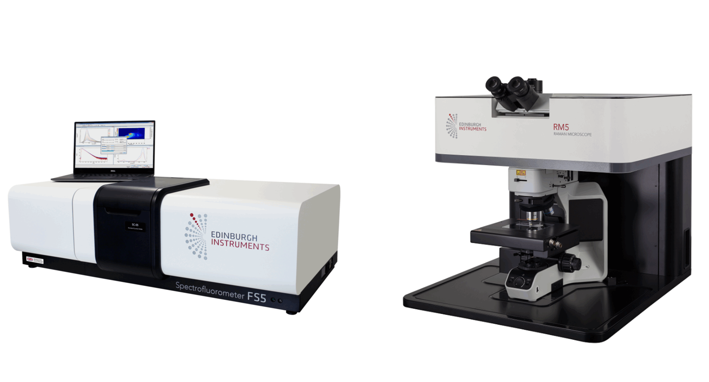
Figure 2. Edinburgh Instruments FS5 Spectrofluorometer and RM5 Confocal Raman Microscope.
The PL probe works through a form of AIE whereby the PCP probe binds to the nanoplastic surface via electrostatic and hydrophobic interactions. This binding restricts PCP’s intramolecular rotation (RIR) and the intramolecular vibrations (RIV), which, in turn, increases the fluorescence emission (Figure 3).
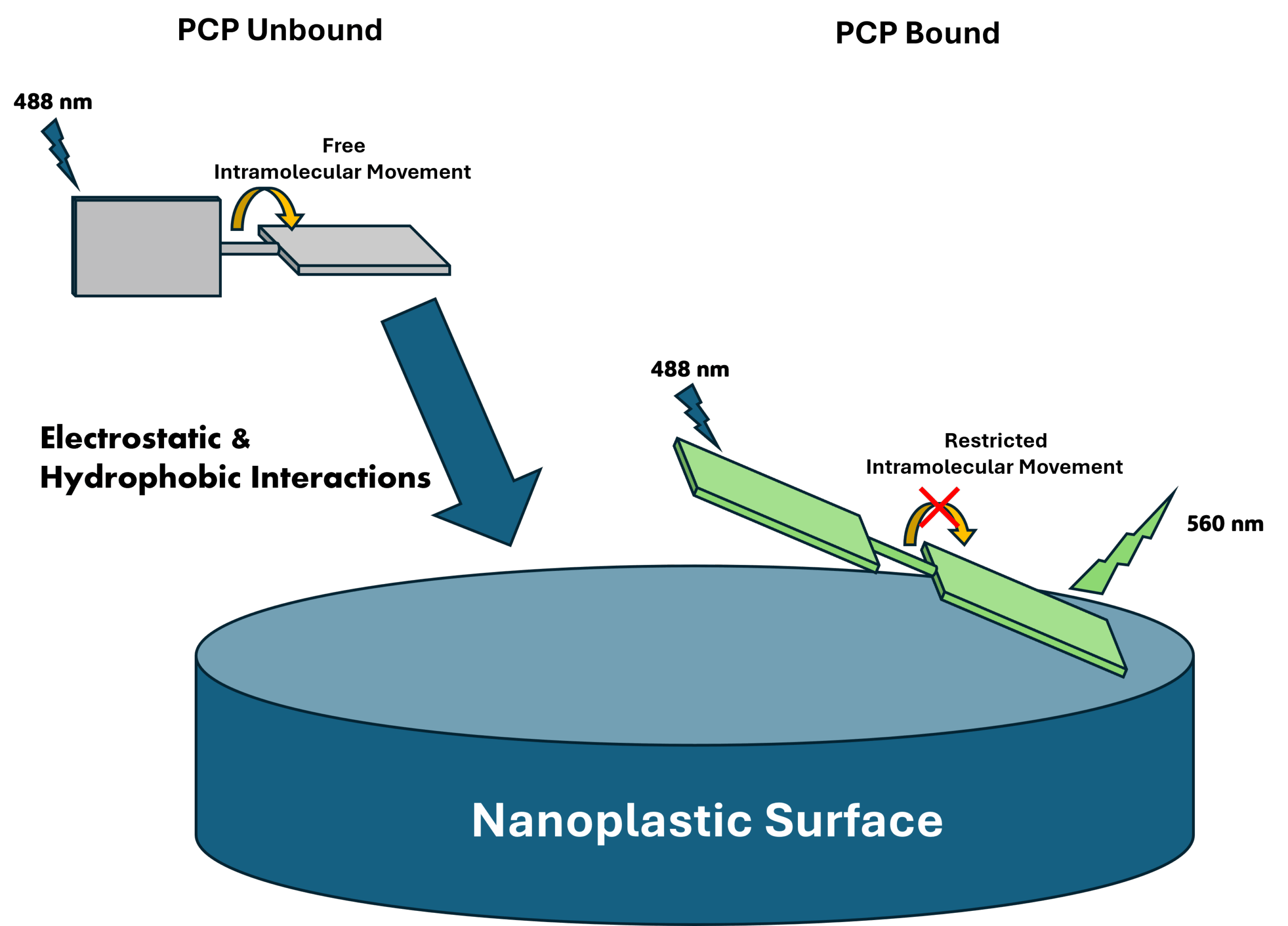
Figure 3. PCP binding to the nanoplastic surface.
Figure 3 shows that restricted intramolecular movement (RIM) upon binding to the nanoplastic stabilises the PCP’s conjugated structure. This inhibits non-radiative decay pathways for excited state electrons, which promotes radiative relaxation, enhancing fluorescence intensity and, in effect, illuminating the particles. These transitions are illustrated in Figure 4.
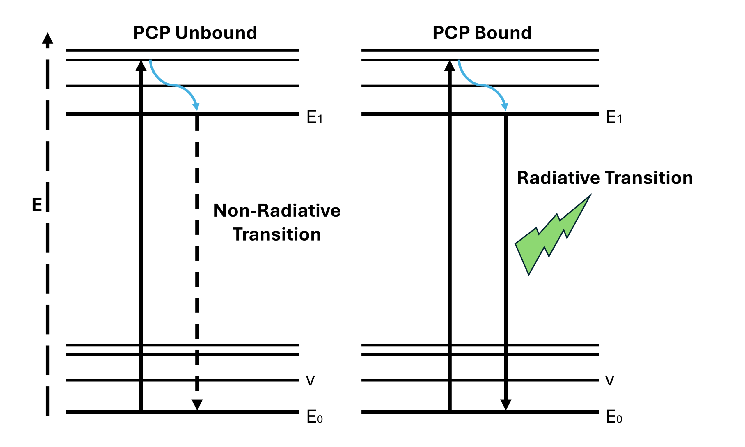
Figure 4. Jablonski diagrams of Unbound PCP and Bound PCP.
Specificity
Once the PCP PL probe had been synthesised, its effectiveness had to be determined. Initially, the fluorescence intensity of the bare PCP was measured before any binding to the nanoplastics. Carboxylation-modified polystyrene (PS-COOH) was then added, and the fluorescence intensity was measured again. PS-COOH was chosen as a model due to its high affinity with PCP. It was found that the fluorescence intensity of PCP significantly increases upon binding to PS-COOH. (This can be seen in Fig.2 A of the original article)
Stability
To determine how effective the PCP probe may be in the long term, the time response properties of the PCP to polystyrene nanoplastics were investigated. To do this, the fluorescence spectrum was acquired at minute intervals for 60 minutes. By comparing the fluorescence intensity of each measurement, the time response can be determined.
The kinetic study demonstrated that there was little to no difference in fluorescence intensity over the course of 60 minutes, displaying the probe’s stability. It also shows that the probe has rapid binding ability to the nanoplastics, with the binding taking place in less than 1 minute. This data is displayed in figure S6 of the supplementary data provided with the original article.
The next step was to determine if the probe could be used for the quantitative detection of nanoplastics. To do this, solutions containing PS-COOH at different concentrations were prepared. The PCP probe was then added to each solution, and the fluorescence intensity was measured. It was observed that the fluorescence intensity increased proportionally with PS-COOH concentration. This is seen in figure 3 of the original article. This highlights the PCP probe’s ability to detect nanoplastics and provide quantitative data on their concentration.
To verify the mechanism by which the PCP probe works, Raman microscopy was used. When performing these measurements, a 785 nm laser was chosen. By choosing a NIR excitation source the researchers could avoid the fluorescence overwhelming the Raman spectrum. This is a fantastic example of how PL and Raman can be used in tandem by careful laser choice.
The Raman spectrum was taken of; pure PCP, pure PS-COOH, PS-COOH@PCP, pure PS-NH2 and PS-NH2@PCP. Binding of PCP to PS-COOH, showed the Raman spectrum changing significantly from the original PCP spectrum. However, when PS-NH2 binds to PCP, the Raman spectrum shows the PCP’s molecular vibrations.
This suggests that upon binding with PCP, PS-COOH restricts the PCP’s intramolecular movement, thereby suppressing its vibrations (RIV), hence the PCP’s peaks cannot be seen. When the PCP is bound to PS-NH2 its intramolecular movement is not restricted. Therefore PCP is free to vibrate, giving the Raman spectrum the same peaks as PCP. This is due to PCP’s low affinity for PS-NH2. These Raman spectra can be viewed in C and D of Figure 2 in the original article.
PCP has a much lower affinity for PS-NH2 than PS-COOH due to the charges at the surface of the modified polystyrene particles. PS-COOH has a negative charge at its surface, this allows for strong electrostatic interactions with the positively charged PCP. PS-NH2 however, has a positively charged surface, making the interactions between the plastic particle and PCP much weaker.
This Research Highlight demonstrates that the PCP probe exhibits high sensitivity and selectivity towards nanoplastics, with a rapid response and excellent stability. Raman Microscopy confirms the mechanism of action, revealing that the increase in fluorescence intensity upon binding to the nanoplastic is due to a restriction in the probe’s intramolecular movement. This new approach is a valuable tool for the selective and quantitative detection of nanoplastics within environmental and biological systems.
The results discussed in this Research Highlight were published in Journal of Hazardous Materials. More details can be found here: https://www.sciencedirect.com/science/article/pii/S0304389423019787?via%3Dihub