Photon antibunching is a signature of single-photon emitters, such as quantum dots, or defect centres in a material. Single-photon emitters are essential for advancing quantum technologies such as quantum networks, communication, and computing, making the characterisation of their antibunching properties increasingly important.
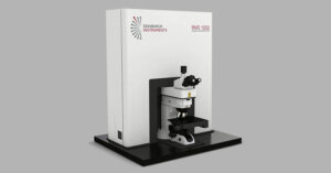
Edinburgh Instruments RMS1000 microscope or FLS1000 with MicroPL upgrade can be configured for antibunching measurements making them ideal for investigating single-photon emitters for quantum applications. In this Application Note, we present the Antibunching Hanbury Brown-Twiss (HBT) Module for the RMS1000 Confocal Multimodal Microscope.
The RMS1000 antibunching upgrade uses a HBT arrangement, shown in Figure 1. The external antibunching HBT module is coupled to the external port of the RMS1000 and incorporates two high–speed hybrid photomultiplier tube detectors (HS-HPD) for coincidence detection. NIR detector options are also available for the antibunching HBT module.
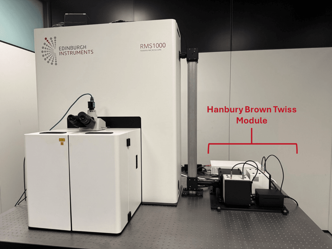
Figure 1. RMS1000 configuration for g(2) correlation measurements showing coupling to antibunching HBT module.
In this setup, light emitted by the sample is split by a 50/50 beamsplitter and sent to two photon-counting detectors. One detector is connected to the Start channel of the time-correlated single photon counting (TCSPC) electronics, and the other one is connected to the Stop channel. The electronics accurately record the delay between two photon events which is registered as counts in a histogram.
Single–photon emitters present a correlation in photon arrival times which creates a dip in the signal histogram, with a shape that depends on the emitter’s lifetime.
g(2) correlation measurements were used to confirm single–photon emission from germanium-vacancy (GeV) centres in diamond, which are prominent, optically active defects used as single-photon sources and quantum emitters.
The sample was prepared by Fondazione Bruno Kessler and consisted of an Element Six electronic-grade diamond (CVD, (100), [N] < 5 ppb) in which germanium ions were implanted with a Poisson-limited spot dose of 65 ions per spot.1 After the implantation, the sample was subjected to a thermal treatment to activate the colour centres.
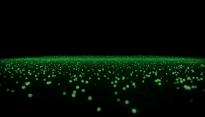
The RMS1000 was equipped with 515 nm CW laser and EMCCD camera and the antibunching HBT module. First, a photoluminescence (PL) map was collected over the surface of the sample using the EMCCD camera. Figure 2A shows the PL intensity of the 602 nm emission showing defined points of high signal intensity.
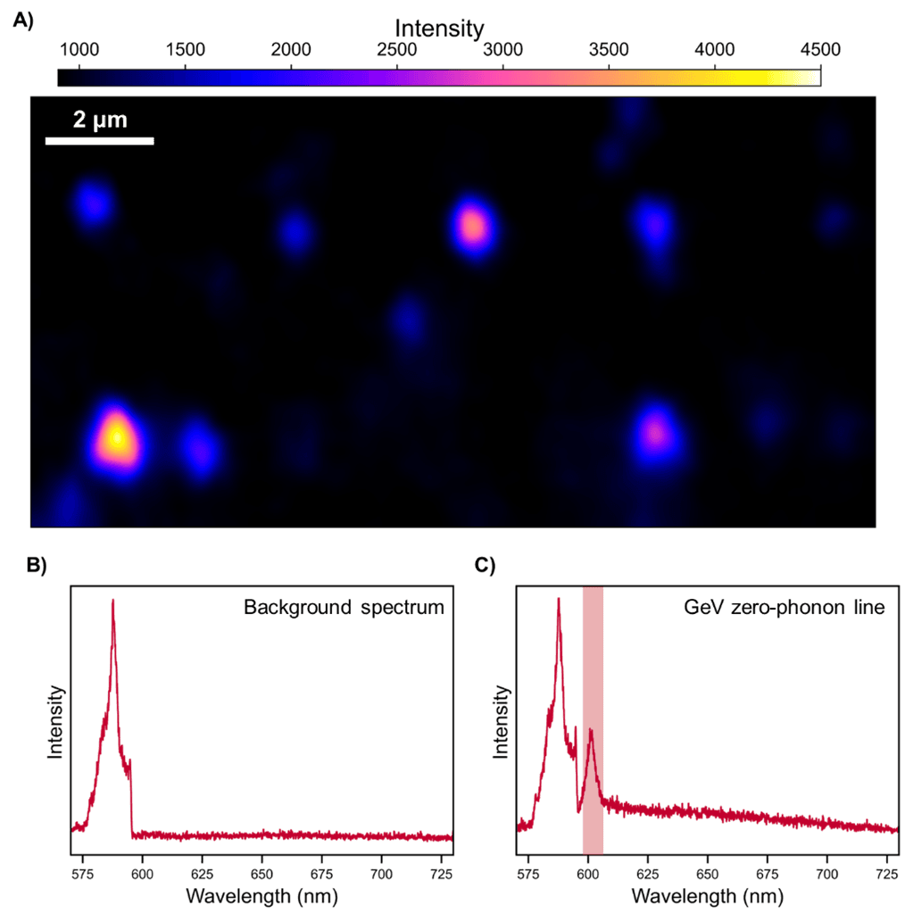
Figure 2. PL analysis of germanium ions implanted in CVD diamond. A) PL intensity map of sample showing intensity of peak at around 602 nm, B) Background spectrum, C) Spectrum of GeV highlighting zero-phonon band at 602 nm.
The background spectrum (Figure 2B) shows only the second-order diamond Raman peaks. The spectrum from a region of high signal intensity (Figure 2C) showed a peak at around 602 nm characteristic of the zero-phonon line (ZPL) of the GeV centre. This peak was absent in background regions, confirming that the GeV PL was spatially localised.
Antibunching measurements were then collected from a region of high PL intensity for verification of single-photon emission. The data was first post-processed to remove the detector background and then g(2) was calculated, the results are shown in Figure 3.
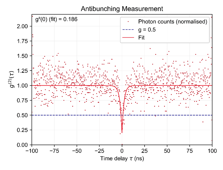
Figure 3. g(2)(τ) plot of Element Six electronic-grade diamond with implanted Germanium ions.
A single emitter cannot emit two photons simultaneously. Measurements of the second-order coherence function, g(2)(τ), show a characteristic “dip” at zero delay (τ = 0). A value of g(2)(0) < 0.5 is the standard benchmark indicating that the emission is single quantum emission rather than from multiple sources. The results show that for this sample the fitted g(2)(0) was 0.186. This result met the <0.5 threshold and confirmed the emission was from a single quantum emitter.
Many defect centres used for quantum networking (e.g., SiV⁻, SnV⁻, telecom emitters) emit in the near infrared (NIR), making measuring NIR antibunching essential. For measurement of these samples, it is possible to couple a NIR detector, such as a NIR–PMT or superconducting nanowire single-photon detector (SNSPD), to the RMS1000.
For antibunching experiments, a SNSPD offers a more sensitive detector option compared to a PMT detector, as it has higher detection efficiency, lower noise and faster timing resolution for NIR measurements (Figure 4).
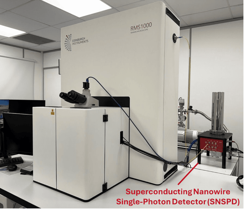
Figure 4. RMS1000 coupled to SNSPD for NIR antibunching experiments.
Upgrading the RMS1000 with a NIR detector enables NIR fluorescence intensity mapping, antibunching, and NIR fluorescence lifetime imaging microscopy (FLIM) applications. This will allow the study of detecting single-photon emission and measuring emitter lifetimes across the NIR and telecom spectral range.
The RMS1000 Confocal Multimodal Microscope can be configured into a powerful platform for single–photon research through visible and NIR antibunching upgrades. Using the Antibunching HBT Module, we demonstrated clear antibunching behaviour from GeV centres in diamond, confirming single–photon emission with g(2)(0)< 0.5.
Additional NIR detection options – including NIR-PMTs and SNSPD coupling – extend these capabilities to emitters with infrared emission, supporting advanced quantum photonics studies such as NIR FLIM, correlation measurements, and high sensitivity mapping.
Together, these upgrades make the RMS1000 and the FLS1000, flexible and scalable tools for quantum material characterisation.
We would like to thank our customers at Fondazione Bruno Kessler for sample fabrication and analysis.
Fabrication of samples: A. Cian, E. Scattolo, and D. Giubertoni.
Sample measurement: E. Missale, E. Nieto Hernandez, and R. Dell’Anna.