Welcome to Edinburgh Instruments monthly blog celebrating our work in Raman, Photoluminescence, and Fluorescence Lifetime Imaging. Every month we will highlight our pick for Map of the Month to show how our spectrometers can be used to reveal all the hidden secrets in your samples.
Two-photon excited fluorescence (2PEF) and second harmonic generation (SHG) are complementary multiphoton imaging techniques for studying biological samples, for example mouse intestine. Mice and humans are similar anatomically, therefore scientific models using mice are frequently applied to human research.
Multiphoton images employ femtosecond pulsed infrared excitation light to generate shorter wavelength light to image the sample. In 2PEF, two infrared photons are simultaneously absorbed by a fluorophore promoting it to an excited state which then radiatively relaxes emitting shorter wavelength fluorescence. In contrast, SHG is not an absorption and emission process and instead the two infrared photons combine in a non-linear optical material with a particular symmetry to generate a new photon with exactly half the wavelength of the incident photons. The versatility of the RMS1000 Raman Microscope allows external lasers to be coupled to the system for multiphoton imaging.

Figure 1: Two-Photon Excited Fluorescence vs Second Harmonic Generation.
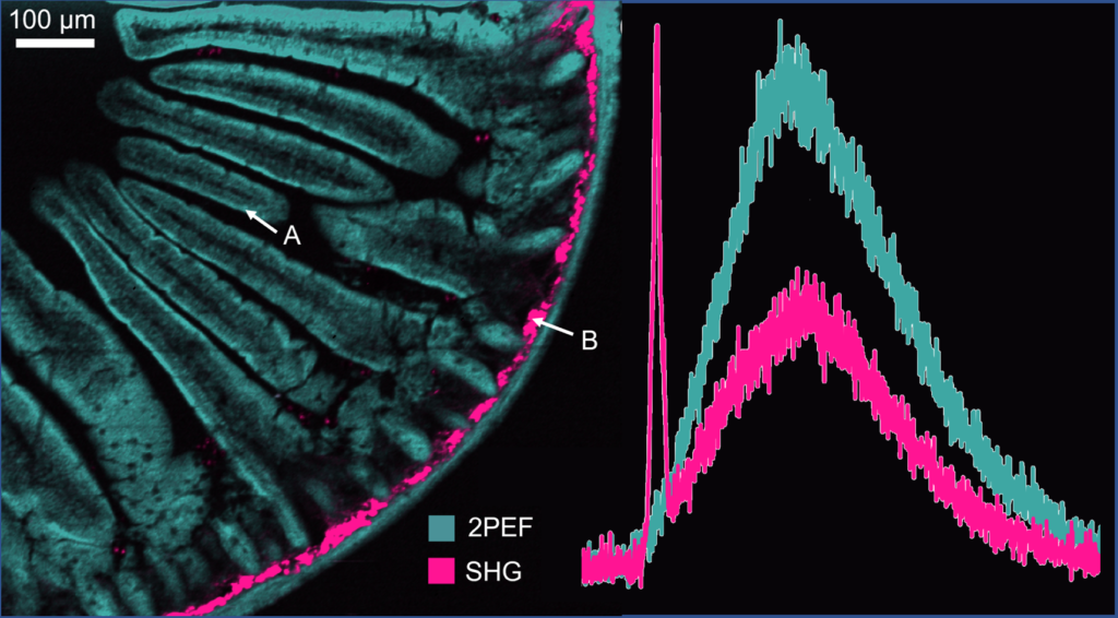
2PEF and SHG signal from the sample can be obtained simultaneously on the CCD camera. The resulting multiphoton image is shown in Figure 2(a). 2PEF at 630 nm from the Alexa Fluor® 568 dye is shown in teal and reveals the structure of the intestinal villi. The area shown in pink is SHG at 520 nm from fibrillar collagen near the intestinal wall. Collagen build up in intestine wall is indicative of many health problems such as Chron’s disease and colon disease. SHG only occurs from molecular structures that are non-centrosymmetric and fibrillar collagen is a common biological structure with this property, eliciting a strong SHG response.
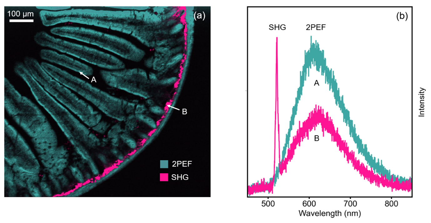
Figure 2: (a) 2PEF and SHG image of mouse intestine section stained with Alexa Fluor® 568 and (b) extracted spectra from two points in the image.
To find out more about how the RMS1000 can benefit your research, contact our sales team today who will be happy to help you.