Skip to content
Skip to content
  • News
  • Events
  • eBooks
  • Blog
  • Careers
  • Contact
  • News
  • Events
  • eBooks
  • Blog
  • Careers
  • Contact
KNOWLEDGEBASE
  • About Us
  • Products

    Fluorescence Spectrometers

    • FLS1000 Photoluminescence Spectrometer
    • FS5 Spectrofluorometer
    • LifeSpec II Lifetime Spectrometer
    • Mini-tau Lifetime Spectrometer

    Raman Microscopes

    • RM5 Raman Microscope
    • RMS1000 Multimodal Confocal Microscope

    Transient Absorption

    • LP980 Transient Absorption Spectrometer

    FTIR Spectrometers

    • IR5 FTIR Spectrometer

    Lasers and LEDs

    • Pulsed Lasers
    • Gas Lasers
    • Customisation Options
    View All Products
  • Techniques
  • Applications
KNOWLEDGEBASE
Edit Content
  • About Us
  • Products
  • Techniques
  • Applications
  • Knowledgebase
  • eBooks
  • News
  • Events
  • Blog
  • Careers
  • Contact Us

RESOURCES

Two-Photon Fluorescence Microscopy with the RMS1000 Confocal Microscope

  • August 9, 2022

KEY POINTS

  • Two-photon microscopy uses two infrared photons to generate shorter wavelength fluorescence.
  • It's ideal for deep imaging of thick biological samples due to localized excitation and lower absorption/scattering of infrared light.
  • This technique can be used for two-photon spectral and lifetime microscopy when coupled with mode-locked femtosecond lasers.
Edit Content

Two-photon microscopy is an imaging technique where two infrared photons are simultaneously absorbed to generate shorter wavelength fluorescence. Two-photon absorption is a non-linear process with the absorption probability scaling quadratically with excitation intensity. This results in the fluorescence being restricted to a much smaller focal volume than in one-photon fluorescence microscopy as the focal point is the only point in space with enough photon density for two-photon absorption to occur. This localised excitation combined with the lower absorption and scattering of the longer wavelength infrared excitation light makes two-photon microscopy ideal for imaging deep into thick biological samples.

Two photon excitation focal volume

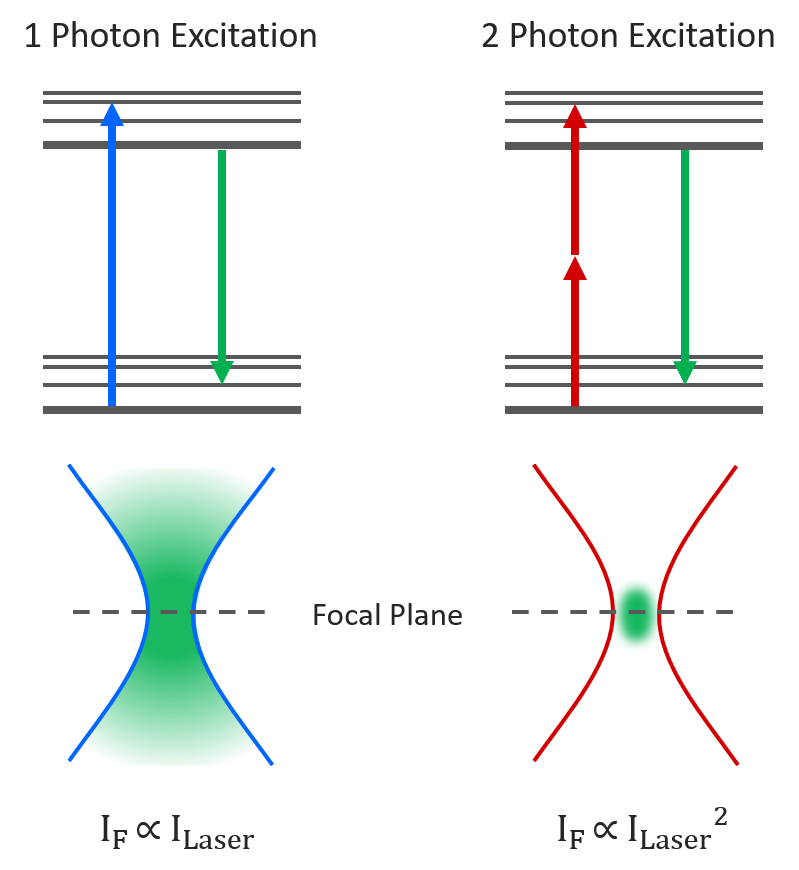
Figure 1: One-photon vs Two-photon Excitation.

Experimental Configuration

Two-photon absorption requires very high excitation intensities which can only be achieved with mode-locked femtosecond pulsed lasers. Traditionally this has been titanium sapphire lasers, but femtosecond fiber lasers are becoming an increasingly attractive and economical alternative. The Edinburgh Instruments RMS1000 Confocal Raman Microscope has external laser coupling ports that enable the optical coupling of mode-locked femtosecond lasers into the microscope. An experimental setup for two-photon spectral and lifetime microscopy with the RMS1000 is shown below, where the RMS1000 has been configured with both a CCD camera and a photon counting Hybrid Photodetector and time-correlated single photon counting (TCSPC) electronics for spectral and lifetime two-photon microscopy.

Two photon microscopy setup

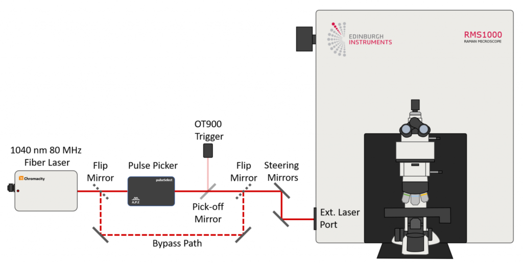
Figure 2: Example optical setup for two-photon microscopy. The RMS1000 is equipped with a BI-CCD camera for spectral fluorescence acquisition and a Hybrid Photodetector and TCSPC lifetime electronics for fluorescence lifetime acquisition. The laser excitation source is a Chromacity 1040 HP femtosecond fiber laser operating at 80 MHz (Chromacity Ltd., UK). For lifetime imaging the output of the laser is pulse picked to the desired pulse frequency using the Pulse Picker pulseSelect (APE GmbH, Germany). A small fraction of the output is picked off into an Edinburgh Instruments OT900 optical trigger to trigger the TCSPC electronics. For spectral measurements the pulse picker is bypassed and the 80 MHz laser output is coupled directly into the RMS1000.

Examples of Two-Photon Imaging with the RMS1000

Kidney Tissue Section

Two photon imaging of mouse kidney cells

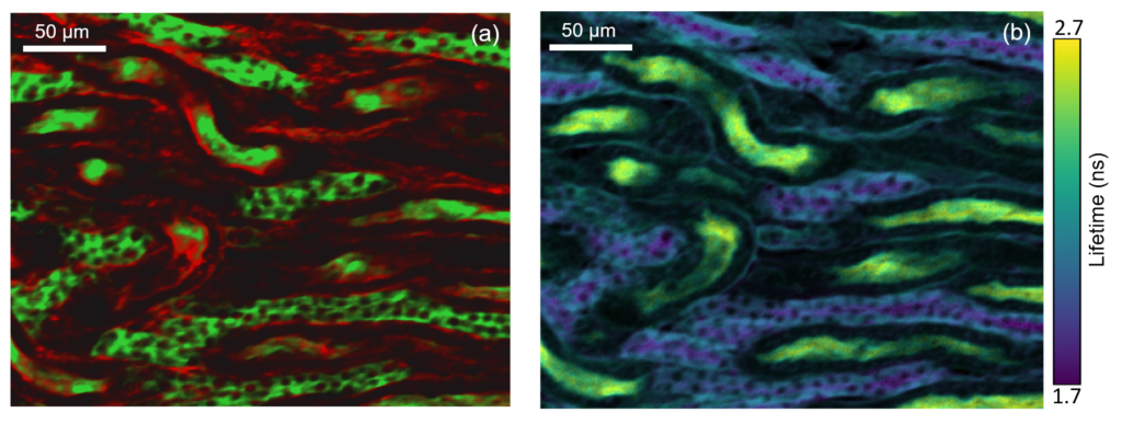
Figure 3: Two photon intensity (a) and lifetime (b) imaging of a mouse kidney section. The glomeruli and convoluted tubules of the kidney tissue were labelled with green fluorescent Alexa Fluor® 488 while the filamentous actin present in glomeruli and the brush border was stained with red-orange fluorescent Alexa Fluor® 568. In (a) the Alexa Fluor® 488 and Alexa Fluor® 568 were excited by the 80 MHz 1040 nm femtosecond laser output and the green and red fluorescence simultaneously captured on the RMS1000 CCD camera. In (b) the 1040 nm laser was pulse picked to 20 MHz and the fluorescence decay measured on the RMS1000 Hybrid Photodetector using TCSPC.

Intestine Tissue Section

Two-photon imaging of mouse intestine

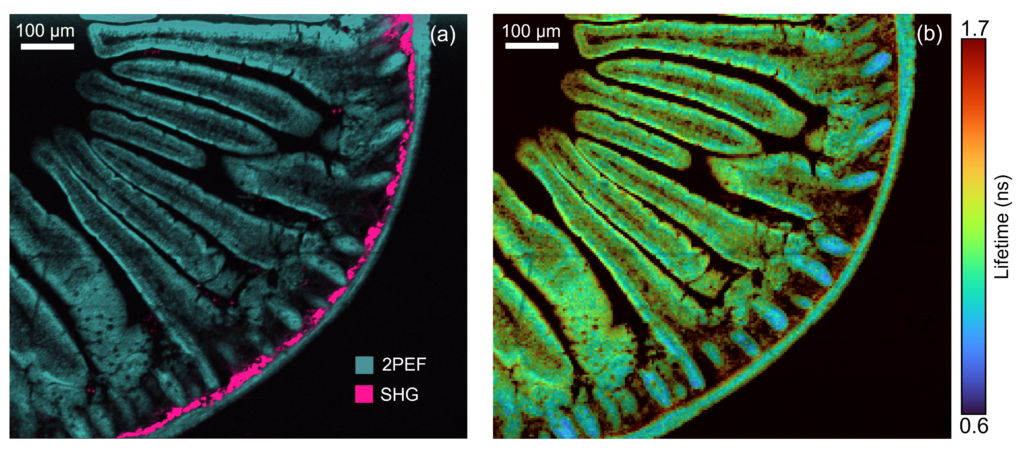
Figure 4: Two photon intensity (a) and lifetime (b) imaging of a mouse intestine section stained with red-orange fluorescent Alexa Fluor® 568. In (a) the Alexa Fluor® 568 was excited by the 80 MHz 1040 nm femtosecond laser output and the two-photon excited fluorescence (2PEF) captured on the RMS1000 CCD camera. Two photon imaging setups can also be used for complementary technique of second harmonic generation (SHG) imaging. SHG at 520 nm from collagen was simultaneously recorded on the RMS1000 CCD camera revealing collagen build up in the intestine wall. In (b) the 1040 nm laser was pulse picked to 20 MHz and the fluorescence decay measured on the RMS1000 Hybrid Photodetector using TCSPC.

Convallaria Rhizome

two photon imaging of convallaria rhizome

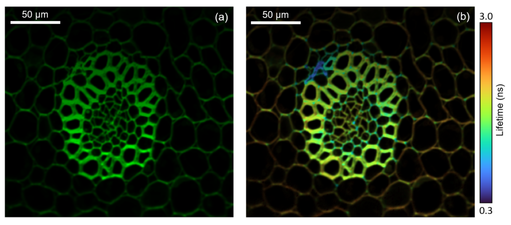
Figure 5: Two photon intensity (a) and lifetime (b) imaging of a convallaria rhizome stained with acridine orange. The acridine orange was excited at 10 MHz using the pulse picked 1040 nm femtosecond laser output and the fluorescence decay measured on the RMS1000’s Hybrid Photodetector using TCSPC. The integrated fluorescence intensity of the decay is shown in (a) and the fitted lifetime in (b), highlighting the increased local environment information that can be obtained from fluorescence lifetime imaging.

RELATED PRODUCTS

RMS1000

Multimodal Confocal Microscope

VIEW

Contact our expert team today to find out more about how our products can improve your research

Contact Us
PrevPrevious
NextNext
Previous Ramacle® Software Highlight: FastMAP® Next Multiphoton Imaging of Mouse Intestine

RESOURCES

Tags:
  • Technical Notes
  • RMS1000
  • Photoluminescence
  • Raman
Technical Note:
Download PDF
Suggested Reading:

Second Harmonic Generation Microscopy with the RMS1000 Confocal Microscope

View more »

Multiphoton Imaging of Mouse Intestine

View more »
Share:

Keep up to date with the latest from Edinburgh Instruments

Join our mailing list and keep up with our latest videos, app notes and more!

LOCATION:
  • Edinburgh Instruments Ltd.
    2 Bain Square, Kirkton Campus, Livingston, EH54 7DQ.
  • sales@edinst.com
  • +44 1506 425 300
ABOUT:
  • About Us
  • Techniques
  • Applications
  • Knowledgebase
  • About Us
  • Techniques
  • Applications
  • Knowledgebase
PRODUCTS:
  • Fluorescence Spectrometers
  • Raman Microscopes
  • UV-Vis Spectrophotometers
  • Transient Absorption
  • FTIR Spectrometers
  • Lasers and LEDs
  • Customisation Options
  • Software
  • Upgrades
  • All Products
  • Fluorescence Spectrometers
  • Raman Microscopes
  • UV-Vis Spectrophotometers
  • Transient Absorption
  • FTIR Spectrometers
  • Lasers and LEDs
  • Customisation Options
  • Software
  • Upgrades
  • All Products
LEGALS:
  • News
  • Events
  • Blog
  • Careers
  • Contact Us
  • Terms and Conditions
  • Privacy Policy
  • News
  • Events
  • Blog
  • Careers
  • Contact Us
  • Terms and Conditions
  • Privacy Policy
SOCIALS:
Youtube Linkedin X-twitter Facebook
©2024 Edinburgh Instruments. Registered in England and Wales No: 962331. VAT No: GB 271 7379 37
Manage Consent
To provide the best experiences, we use technologies like cookies to store and/or access device information. Consenting to these technologies will allow us to process data such as browsing behaviour or unique IDs on this site. Not consenting or withdrawing consent, may adversely affect certain features and functions.
Functional Always active
The technical storage or access is strictly necessary for the legitimate purpose of enabling the use of a specific service explicitly requested by the subscriber or user, or for the sole purpose of carrying out the transmission of a communication over an electronic communications network.
Preferences
The technical storage or access is necessary for the legitimate purpose of storing preferences that are not requested by the subscriber or user.
Statistics
The technical storage or access that is used exclusively for statistical purposes. The technical storage or access that is used exclusively for anonymous statistical purposes. Without a subpoena, voluntary compliance on the part of your Internet Service Provider, or additional records from a third party, information stored or retrieved for this purpose alone cannot usually be used to identify you.
Marketing
The technical storage or access is required to create user profiles to send advertising, or to track the user on a website or across several websites for similar marketing purposes.
Manage options Manage services Manage {vendor_count} vendors Read more about these purposes
View preferences
{title} {title} {title}