Resonance Raman spectroscopy is a Raman enhancement technique in which the laser excitation frequency is chosen to be close to the frequency of an electronic transition of the sample. Resonance Raman can enhance the Raman scattering intensity by a factor of 102-106 and improves signal-to-noise. The enhanced Raman scattering means shorter exposure times can be used, allowing much faster spectral acquisition times. Additionally, samples at extremely low concentrations can easily be studied.
When the laser frequency exactly matches an electronic transition of the sample it is said it be in resonance, shown in Figure 1. This is where the greatest enhancement occurs and hence tuneable lasers are the ideal excitation source for resonance Raman spectroscopy. However, as long as the laser wavelength lies close enough to the electronic transition, enhancement will still be observed.1 Therefore the only requirement for a Raman system to study resonance Raman scattering is an excitation laser near the correct frequency. The Edinburgh Instruments RM5 Raman Microscope can fully integrate up to three lasers at one time; therefore, making it is easy to select three different excitation wavelengths that are close to the resonance of your samples.
Resonance Raman spectroscopy can be thought of as selective, only the part of the sample with the highest light absorption will give enhancement, termed the chromophore. Furthermore, just the part of the molecule associated with the chromophore will have its Raman scattering intensities enhanced. This also simplifies the Raman spectra a great deal, which can make interpretation easier. A chromophore is a chemical group responsible for giving molecules their colour. Thus, resonance Raman is useful for analysis coloured samples.
One drawback of Resonance Raman spectroscopy is increased fluorescence which can swamp the Raman signal. When you are matching the laser to an absorption band, absorption will occur meaning there is a high chance of fluorescence obscuring the Raman peaks. Figure 1 highlights the reason why fluorescence is more of an issue for Resonance Raman compared to standard Raman scattering. The interference of fluorescence limits the number of molecules studied by Resonance Raman spectroscopy. Pre-resonance can also be used, Figure 1, this is where the laser excitation is around 100 wavenumbers below the electronic transition. Here some enhancement can still be seen but with a lower risk of fluorescence.
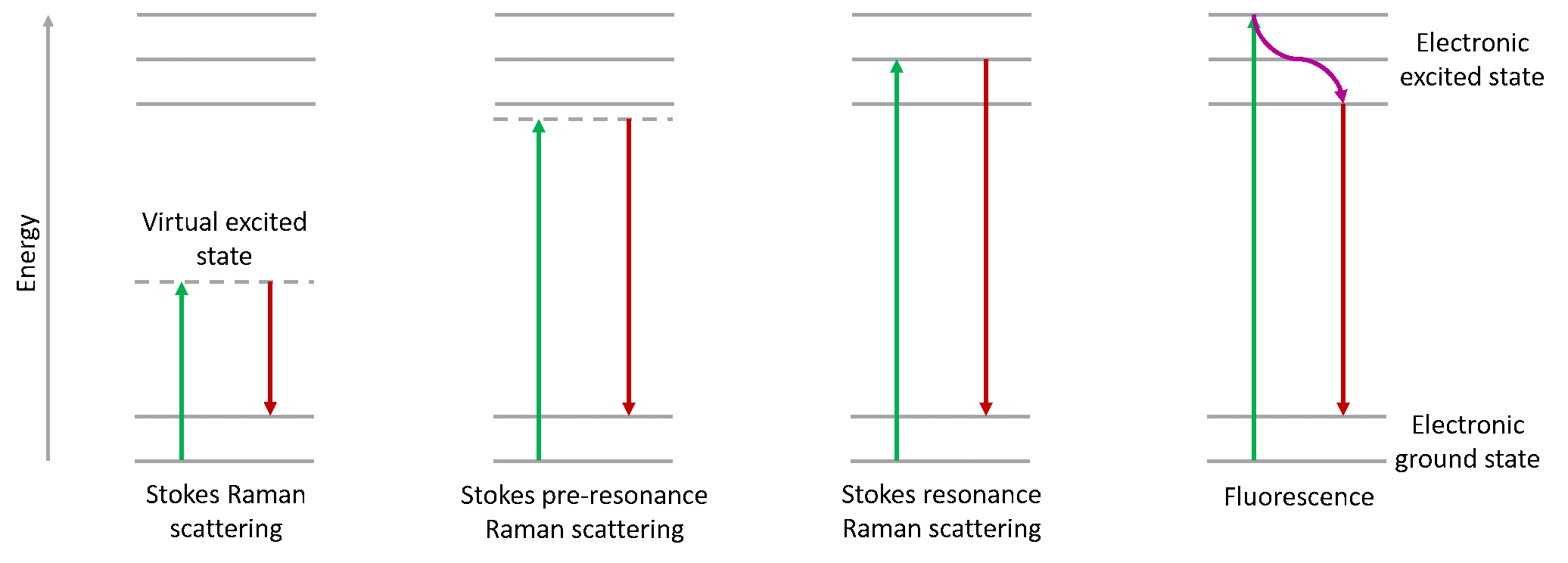
Figure 1: Energy diagram showing Raman scattering, pre-resonance Raman scattering, resonance Raman scattering, and fluorescence.
Resonance Raman spectroscopy is popularly used for the analysis of biological materials in the UV region. UV-Resonance Raman spectroscopy in the deep UV is particularly useful for the study of nucleic acids, such as DNA and RNA. Proteins and peptides are also well studied in the UV for structural and folding information. In the visible region Resonance Raman has provided in depth data on haem groups, carotenoids, and pigments.2 Overall Resonance Raman spectroscopy is an easy technique to carry out if the Raman system houses the correct laser excitation wavelength, and sample fluorescence and damage effects are minimal.
You can find out more about our RM5 Raman Microscope on our website. If you have enjoyed reading this article and wish to learn more about our applications and products then why not contact a member of our team at sales@edinst.com or follow us on social media via your preferred channel below.
1. Smith, E. & Dent, G. Modern Raman Spectroscopy. Modern Raman Spectroscopy (2019)