
Skip to content
  • News
  • Events
  • eBooks
  • Blog
  • Careers
  • Contact
  • News
  • Events
  • eBooks
  • Blog
  • Careers
  • Contact
KNOWLEDGEBASE
  • About Us
  • Products

    Fluorescence Spectrometers:

    • FLS1000 Photoluminescence Spectrometer
    • FS5 Spectrofluorometer
    • LifeSpec II Lifetime Spectrometer
    • Mini-tau Lifetime Spectrometer

    Raman Microscopes:

    • RM5 Raman Microscope
    • RMS1000 Multimodal Confocal Microscope

    Transient Absorption:

    • LP980 Transient Absorption Spectrometer

    FTIR Spectrometers:

    • IR5 FTIR Spectrometer

    Lasers and LEDs:

    • Pulsed Lasers
    • Gas Lasers

    Options:

    • Upgrades
    • Customisation
    View All Products
  • Techniques
  • Applications
KNOWLEDGEBASE
  • About Edinburgh Instruments
  • Products
  • Techniques
  • Applications Old
  • Knowledgebase
  • eBooks
  • News
  • Events
  • Blog
  • Careers
  • Contact Us

RESOURCES

2D Materials and Multimodal Microscopy: Raman, Photoluminescence and SHG

  • April 24, 2025

KEY POINTS

  • Raman spectroscopy probes lattice vibrations to reveal layer number, defects, strain, doping and structural variations in 2D materials.
  • Photoluminescence (PL) spectroscopy probes electronic structure and band gap, providing insight into layer thickness, defects, strain and the local environment.
  • Second harmonic generation (SHG) imaging is highly sensitive to crystal symmetry and orientation, providing complementary information about stacking and structure.
  • Multimodal microscopy combines Raman, PL, and SHG to provide a more complete picture of the structural, electronic and defect properties of 2D materials than any single technique alone.

Two-dimensional materials have remarkable structural, electronic and optical properties, but understanding how these properties vary across an individual flake requires more than a single measurement.

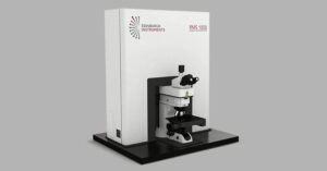
In this Edinburgh Instruments webinar, Angela Flack and Dr Nikesh Patel demonstrate how multimodal microscopy combines Raman spectroscopy, PL and SHG imaging to investigate 2D materials from complementary perspectives. Using the RMS1000 Raman Microscope, the webinar moves from the fundamentals of each technique to live mapping and detailed analysis of molybdenum disulfide and tungsten diselenide samples.


Raman Spectroscopy: Mapping Phonons

Raman spectroscopy provides a vibrational fingerprint of a 2D material by measuring the frequency shifts produced by inelastic scattering. These vibrational modes are highly sensitive to the material’s structure, enabling Raman mapping to investigate layer number, defects, strain, doping and functionalisation across a sample.

During the live demonstration, Raman mapping of molybdenum disulfide reveals clear differences across individual flakes. The webinar also demonstrates fast mapping, reducing the example Raman map from approximately six minutes with standard mapping to around a minute and a half.


Photoluminescence: Probing Electronic Structure

While Raman spectroscopy probes phonons, photoluminescence spectroscopy probes electronic structure and the band gap. Changes in PL emission energy and intensity can reveal variations in layer thickness, defects, strain and interactions with the surrounding environment.

This makes PL imaging particularly valuable for visualising spatial variations across 2D materials. In the molybdenum disulfide example, regions of lower PL intensity are associated with greater layer number, while shifts in PL peak position can indicate potential strain within the flake.


SHG Imaging: Revealing Crystal Symmetry

Second harmonic generation takes the analysis a step further by probing crystal symmetry. SHG is a nonlinear optical process that occurs in non-centrosymmetric materials, making its intensity and polarisation highly sensitive to crystal orientation. This enables SHG imaging to investigate crystal stacking, orientation, domains, grain boundaries and defects in 2D materials.

In the live demonstration, SHG mapping reveals distinct domains and grain boundaries that are less apparent in the corresponding Raman and PL maps.


Building the Complete Picture

The power of multimodal microscopy becomes particularly clear in the analysis of tungsten diselenide. Raman imaging distinguishes monolayer and multilayer regions, while PL spectral analysis enables regions to be assigned as monolayer, bilayer and trilayer. SHG then adds information about crystal symmetry and stacking.

Together, these complementary techniques connect vibrational structure, electronic properties and crystal symmetry within the same sample, providing researchers with a more comprehensive understanding of their 2D materials. This information can help assess synthesis, identify areas for improvement and support more complete material characterisation.


Watch the Multimodal Microscopy Webinar

Watch the full webinar to see Angela Flack and Dr Nikesh Patel showcase Raman, photoluminescence and SHG imaging in action on the RMS1000 Raman Microscope, and discover how combining complementary spectroscopic techniques can uncover more information from 2D materials.

Read More Read Less

RELATED PRODUCTS

RMS1000

Multimodal Confocal Microscope

VIEW

Contact our expert team today to find out more about how our products can improve your research

Contact Us
PrevPrevious
NextNext
Previous Improving Axial Resolution and Spectral Sensitivity in 3D Confocal Raman Imaging Using an Immersion Objective Lens Next What is Radioluminescence?

RESOURCES

Multimodal Microscopy Webinar Featured Image
Tags:
  • Webinars
  • 2D Materials
  • Semiconductors
  • RMS1000
  • Photoluminescence
  • Raman
Featured Product:

RMS1000 Raman Microscope

View more »
Suggested Reading:

High-Resolution Raman & PL Imaging of MoS2

View more »

Multimodal Raman, Photoluminescence & SHG Imaging of CVD Grown WSe2

View more »
Share:

Keep up to date with the latest from Edinburgh Instruments

Join our mailing list and keep up with our latest videos, app notes and more!

LOCATION:
  • Edinburgh Instruments Ltd.
    2 Bain Square, Kirkton Campus, Livingston, EH54 7DQ
  • sales@edinst.com
  • +44 1506 425 300
ABOUT:
  • About Edinburgh Instruments
  • Techniques
  • Applications
  • Knowledgebase
  • About Edinburgh Instruments
  • Techniques
  • Applications
  • Knowledgebase
PRODUCTS:
  • Fluorescence Spectrometers
  • Raman Microscopes
  • UV-Vis Spectrophotometers
  • Transient Absorption
  • FTIR Spectrometers
  • Lasers and LEDs
  • Customisation Options
  • Software
  • Upgrades
  • All Products
  • Fluorescence Spectrometers
  • Raman Microscopes
  • UV-Vis Spectrophotometers
  • Transient Absorption
  • FTIR Spectrometers
  • Lasers and LEDs
  • Customisation Options
  • Software
  • Upgrades
  • All Products
LEGALS:
  • News
  • Events
  • Blog
  • Careers
  • Distribution & Agents
  • Contact Us
  • Terms and Conditions
  • Privacy Policy
  • Coordinated Vulnerability Disclosure Policy
  • News
  • Events
  • Blog
  • Careers
  • Distribution & Agents
  • Contact Us
  • Terms and Conditions
  • Privacy Policy
  • Coordinated Vulnerability Disclosure Policy
SOCIALS:
Youtube Linkedin X-twitter Facebook
©2026 Edinburgh Instruments. Registered in England and Wales No: 962331. VAT No: GB 271 7379 37
Manage Consent
To provide the best experiences, we use technologies like cookies to store and/or access device information. Consenting to these technologies will allow us to process data such as browsing behaviour or unique IDs on this site. Not consenting or withdrawing consent, may adversely affect certain features and functions.
Functional Always active
The technical storage or access is strictly necessary for the legitimate purpose of enabling the use of a specific service explicitly requested by the subscriber or user, or for the sole purpose of carrying out the transmission of a communication over an electronic communications network.
Preferences
The technical storage or access is necessary for the legitimate purpose of storing preferences that are not requested by the subscriber or user.
Statistics
The technical storage or access that is used exclusively for statistical purposes. The technical storage or access that is used exclusively for anonymous statistical purposes. Without a subpoena, voluntary compliance on the part of your Internet Service Provider, or additional records from a third party, information stored or retrieved for this purpose alone cannot usually be used to identify you.
Marketing
The technical storage or access is required to create user profiles to send advertising, or to track the user on a website or across several websites for similar marketing purposes.
  • Manage options
  • Manage services
  • Manage {vendor_count} vendors
  • Read more about these purposes
View preferences
  • {title}
  • {title}
  • {title}