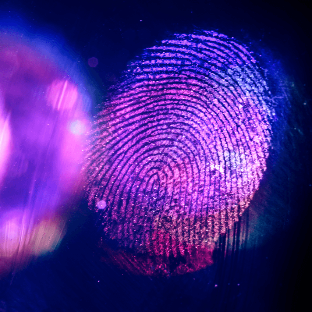
Among the many features that distinguish individuals, fingerprints are uniquely personal, which is why they have been essential to forensic identification for decades. Over the years, a wide range of techniques has been developed to extract and analyse these intricate patterns. Pushing the boundaries in this field, research led by Professor Gangfeng Ouyang and Professor Huan Pang, published at Nature communications under the title “Luminescent nanofibers for human skin textures photocopying”, introduces a cutting‑edge method for high‑resolution optical analysis of human skin textures, opening new possibilities for forensic science and material innovation.
The authors introduced an innovative nanofibre‑based imaging technique (NFIT) that combines fluorescent nanocrystals with electrospinning technology to create a highly sensitive material capable of photocopying detailed human skin textures without any physical contact. Using advanced optical imaging methods, the technique reveals the intricate, micrometre‑scale ridge patterns of human fingerprints, delivering high fidelity and consistent performance even under changing environmental conditions.
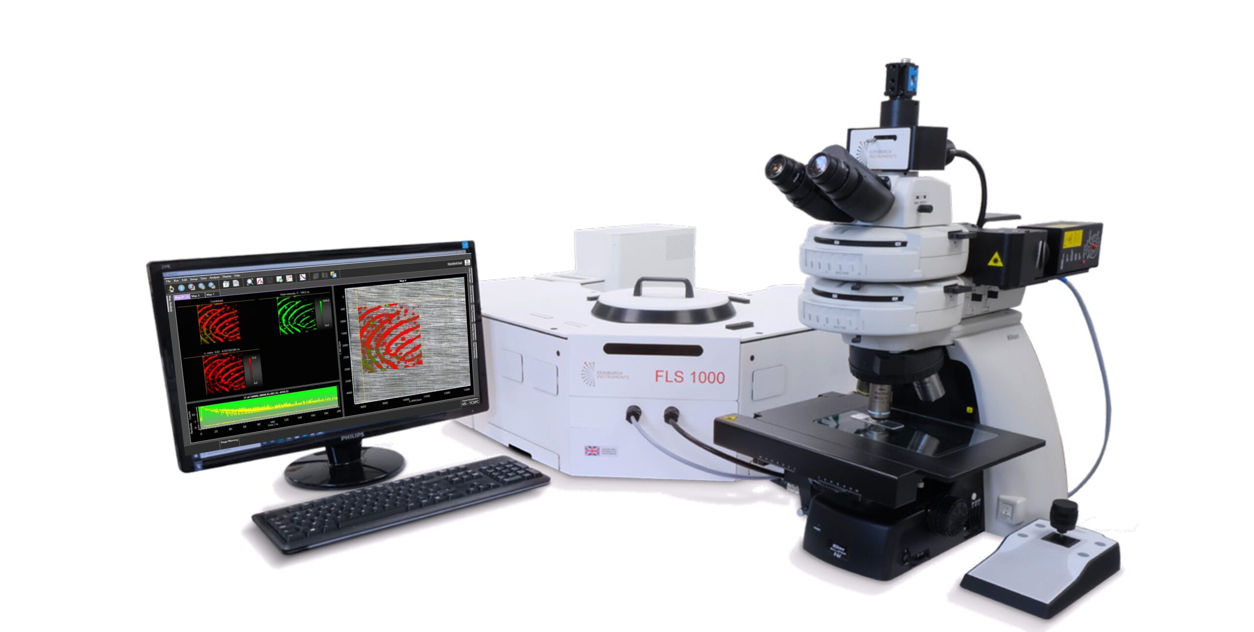
Figure 1. FLS1000 Photoluminescence Spectrometer with an MicroPL upgrade.
Central to this technique are CsPbBr3@HPβCD nanocrystals, which were electrospun together with thermoplastic polyurethane (a common polymer) to produce flexible nanofibres. When these fibres come into contact with fingerprint residues on common surfaces, such as tinfoil, quartz, iron, glass or plastic, they readily adhere to the residues, creating a photocopy of the fingerprint. Under appropriate light excitation, the nanocrystal fibres emit an intense fluorescent signal that sharply delineates the fingerprint structures. The resulting high-contrast images reveal fine detail, help distinguish genuine fingerprints from forged ones, and may also indicate the pressure applied during contact, adding further potentially valuable forensic information.
A detailed characterization, including analytical, morphological and spectroscopical analyses was conducted to clarify howCsPbBr3@HPβCD nanofibres interact with chemical species commonly present in sweat residues more specifically, inorganic salts and organic compounds, including sodium chloride (NaCl), magnesium chloride (MgCl₂), potassium chloride (KCl), and glucose. Photophysical characterisation, encompassing steady‑state, time‑resolved, and temperature‑dependent photoluminescence (PL) measurements, were performed with an FLS1000 Photoluminescence Spectrometer and revealed how these sweat components modulate fluorescence performance and influence fingerprint image quality. Together, these insights highlighted the effectiveness, robustness, and forensic relevance of the NFIT approach.
Going beyond conventional characterisation, the team also conducted in situ Fluorescence Lifetime Imaging Microscopy (FLIM) by using the MicroPL upgrade for the FLS1000 (Figure 2). FLIM is a powerful imaging technique that extends conventional PL imaging by exploiting variations in fluorescence decay rates as an additional contrast mechanism.
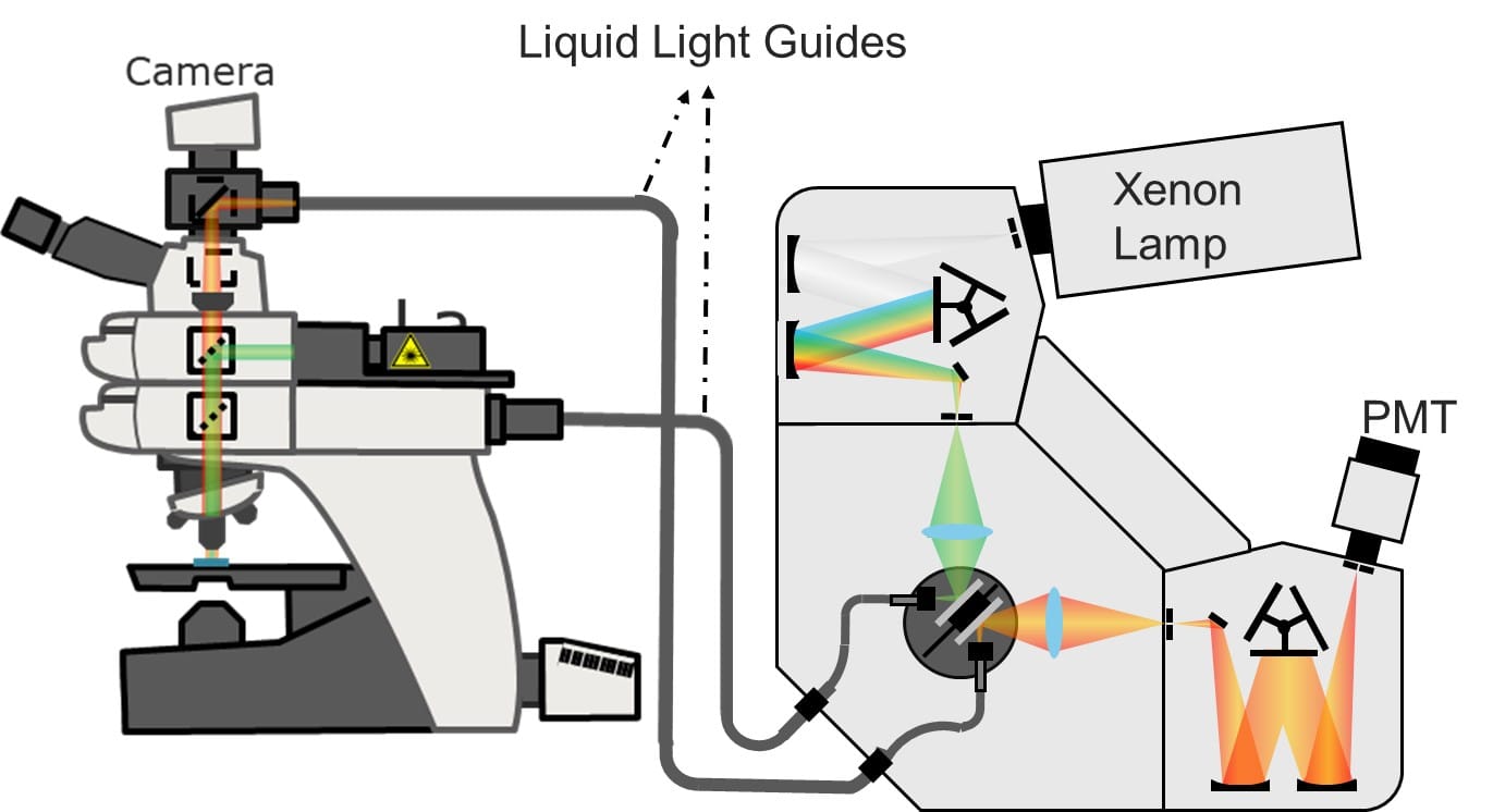
Figure 2. Schematics of the MicroPL microscope upgrade to an FLS1000.
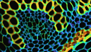
By mapping fluorescence lifetimes across the sample and analysing their spatial variations, the authors confirmed the optical behaviour of the CsPbBr3@HPβCD nanofibres over the imaging area. The technique enabled accurate reconstruction of skin surface features (Figure 3) and revealed that chlorides present in human sweat are predominantly concentrated around the outer edges of sweat pores on the fingerprint ridges. In these regions, the emission exhibited a pronounced blueshift and a shortened fluorescence lifetime (Figure 4), indicating localised chemical interactions, which are an important consideration for more accurate data analysis.
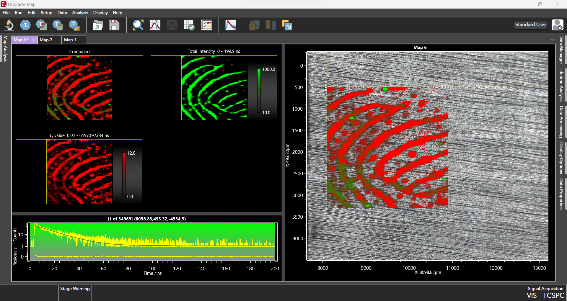
Figure 3. FLIM measurements were acquired and analysed using the FluoracleMap software provided by Edinburgh Instruments. The maps illustrate the total emission intensity and lifetime range for each sample.
The researchers also found that regions with strong emission intensity and relative lifetimes exceeding 100% correlate with glucose concentrations. This finding suggests that the technique could also be used to map glucose distribution and estimate glucose levels, marking a significant advance with promising implications for biomedical diagnostics. They further demonstrated that skin textures from diverse body regions (including the forehead, backs of the hands, and soles of the feet) can also be successfully captured, highlighting the potential of the non‑invasive technique for clinical diagnostics and health monitoring.
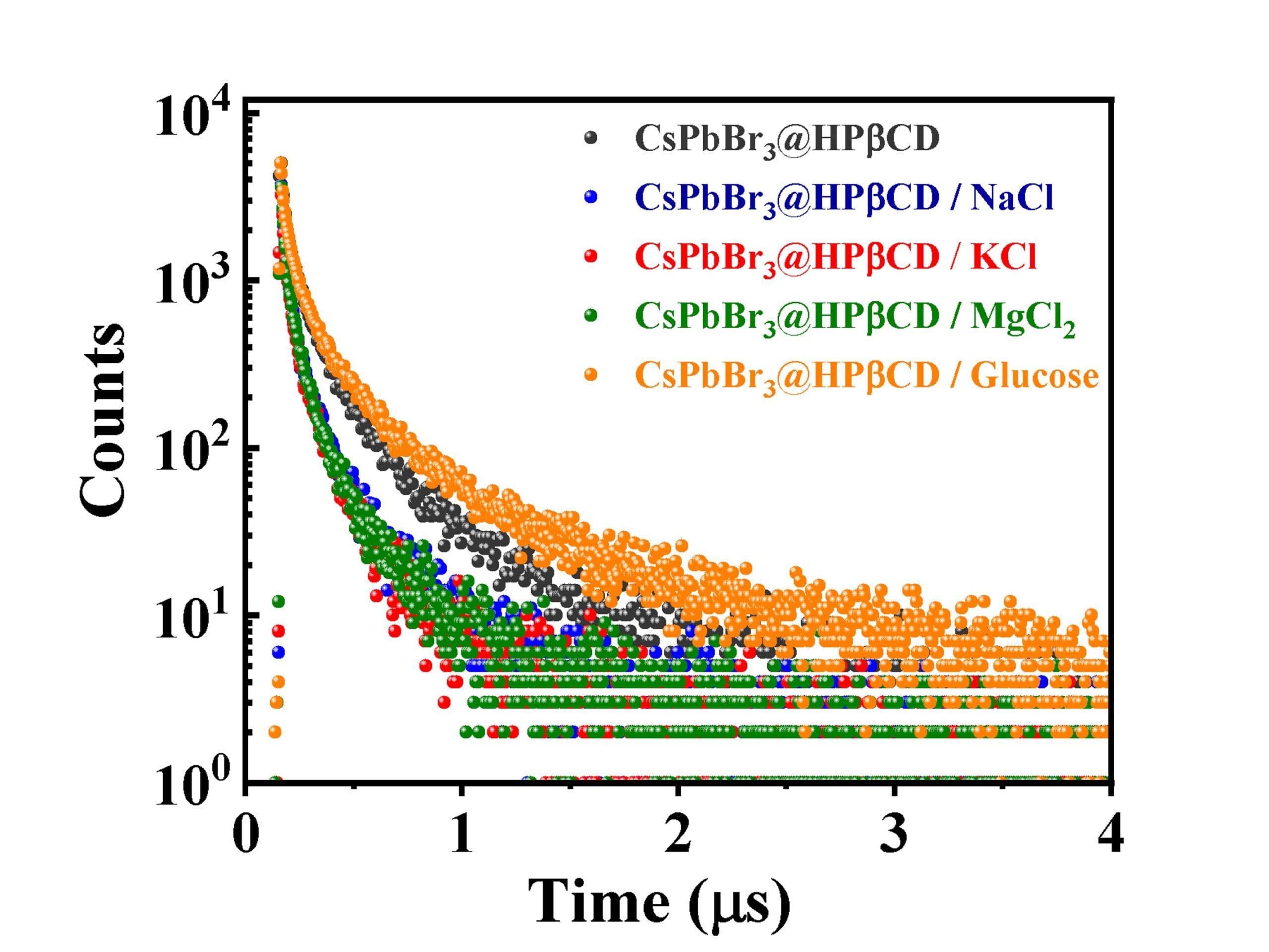
Figure 4. Fluorescence lifetime decay for the neat CsPbBr3@HPβCD nanofibres, and the fibres exposed to selected sweat components: Sodium chloride (NaCl); Potassium chloride (KCl); Magnesium chloride (MgCl2); and Glucose.
To summarize, the MicroPL upgrade was successfully used to characterise fluorescent fingerprints by analysing differences in emission wavelength and lifetime across different regions of the fingerprint, with each region showing stronger interactions with chemical species present in sweat.
We sincerely thank the authors for kindly providing the data and the screenshot of the sample being measured, which greatly supported the preparation of this research highlight.
The full article was published in Nature Communications and can be found at DOI: 10.1038/s41467-025-64703-5.