Skip to content
Skip to content
  • News
  • Events
  • eBooks
  • Blog
  • Careers
  • Contact
  • News
  • Events
  • eBooks
  • Blog
  • Careers
  • Contact
KNOWLEDGEBASE
  • About Us
  • Products

    Fluorescence Spectrometers

    • FLS1000 Photoluminescence Spectrometer
    • FS5 Spectrofluorometer
    • LifeSpec II Lifetime Spectrometer
    • Mini-tau Lifetime Spectrometer

    Raman Microscopes

    • RM5 Raman Microscope
    • RMS1000 Multimodal Confocal Microscope

    Transient Absorption

    • LP980 Transient Absorption Spectrometer

    FTIR Spectrometers

    • IR5 FTIR Spectrometer

    Lasers and LEDs

    • Pulsed Lasers
    • Gas Lasers
    • Customisation Options
    View All Products
  • Techniques
  • Applications
KNOWLEDGEBASE
Edit Content
  • About Us
  • Products
  • Techniques
  • Applications
  • Knowledgebase
  • eBooks
  • News
  • Events
  • Blog
  • Careers
  • Contact Us

RESOURCES

What is Raman Imaging?

  • June 10, 2024

KEY POINTS

  • Raman imaging, also called Raman mapping, measures the variation in the Raman spectrum across a sample.
  • Raman imaging can be performed across a two-dimensional plane, a three-dimensional volume, or undulating or tilted sample surfaces.
  • Raman imaging allows for material identification, quantitative analysis, the detection of strain and crystallinity, layer number in quantum materials, and spectral similarities based on chemometrics.
Edit Content

What is Raman Imaging? 

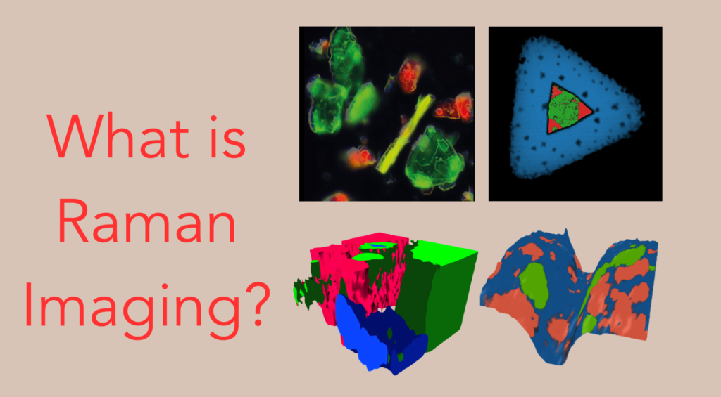
Raman imaging, also called Raman mapping, is a hyperspectral technique where variations in the Raman spectrum across or throughout a sample create a contrast, Figure 1. In a Raman image, each point contains a full Raman spectrum, which is a source of a vast amount of chemical information. False colour images are created from variations in the Raman spectra that allow for spatial information about the chemical composition of the sample to be obtained.  

Raman images

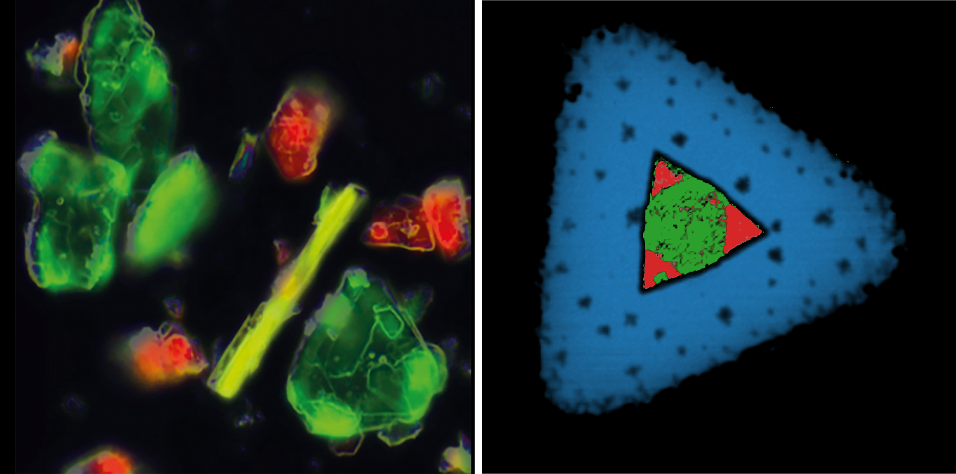
Figure 1. Raman images recorded on Edinburgh Instruments RM5 and RMS1000 Confocal Microscopes. Left: amino acids, and Right: tungsten diselenide crystal. 


How to Record Raman Images 

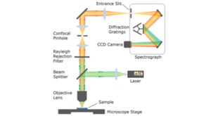
Raman images are acquired using a confocal Raman microscope, Figure 2. The microscope incorporates a laser excitation source, and the beam is focused onto a small spot on the sample using an objective lens. The Raman scattered photons from the sample pass back into the objective and are guided onto a CCD detector.  

confocal raman microscope

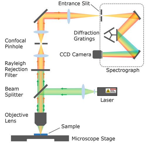
Figure 2. Raman Confocal Microscope. 


Imaging is achieved by a motorised microscope stage that can move in X, Y and Z. Images can be recorded by mapping the sample across the X, Y-plane (Figures 1 and 3), throughout an X, Y, and Z-volume (Figure 4) and in X, Y, and Z across an undulating or tilted surface (Figure 4). Each point where a spectrum is recorded is a pixel in the resulting image.  

Raman imaging technique

Figure 3. 2D Raman imaging methodology.  

3D Raman imaging

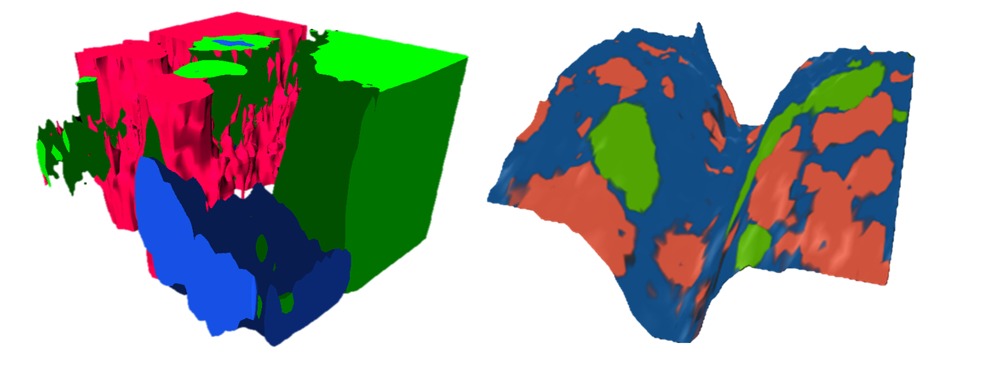
Figure 4. Volumetric Raman image of a geological sample obtained using 3D mapping (left) and surface Raman image of a pharmaceutical tablet using SurfMAP® in Edinburgh Instruments Ramacle software (right).  


What Can Raman Imaging Tell Us?  

Multiple parameters from the spectra recorded at each pixel can be used in constructing a Raman image, Figure 5.  The main aim of Raman imaging is usually to identify the presence of a material and track its distribution across a sample surface or throughout a volume within the sample. This is possible because different materials have unique Raman spectra that can be used as molecular fingerprints for identification. Since Raman bands are narrow and specific to particular vibrational modes, multiple bands and materials can be imaged simultaneously on a sample. Images can be built using the intensity, position, and width of Raman bands, which can track material concentrations, strain, and crystallinity, respectively. Functions that score spectra based on similarities using chemometric techniques can also be used in Raman imaging. In 2D quantum materials like graphene, peak intensity ratios can be used to image layer number, Figure 6. 


Raman spectroscopy

Figure 5. The information that can be obtained from Raman imaging.   

silicon and graphene raman images

Figure 6. Strain image of silicon built using peak position analysis (left) and graphene layer image made using ratiometric analysis (right).    


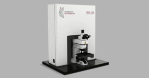
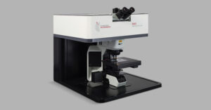
If you would like further information on how the RM5 and RMS1000s Raman Microscope can be used to help you with your research, contact us. 

If you have enjoyed this blog and want to keep up to date with our Raman images, sign up for our monthly eNewsletter below for further updates on our latest research, application notes and products. 

RELATED PRODUCTS

RMS1000

Multimodal Confocal Microscope

VIEW

RM5

Raman Microscope

VIEW

Contact our expert team today to find out more about how our products can improve your research

Contact Us
PrevPrevious
NextNext
Previous What is Photoinduced Electron Transfer? Next Raman and Photoluminescence Characterisation of GaN

RESOURCES

Tags:
  • Spectral School
  • RM5
  • RMS1000
  • Raman
Featured Product:

RMS1000 Raman Microscope

View more »

RM5 Raman Microscope

View more »
Suggested Reading:

What is Confocal Raman Microscopy?

View more »
Share:

Keep up to date with the latest from Edinburgh Instruments

Join our mailing list and keep up with our latest videos, app notes and more!

LOCATION:
  • Edinburgh Instruments Ltd.
    2 Bain Square, Kirkton Campus, Livingston, EH54 7DQ.
  • sales@edinst.com
  • +44 1506 425 300
ABOUT:
  • About Us
  • Techniques
  • Applications
  • Knowledgebase
  • About Us
  • Techniques
  • Applications
  • Knowledgebase
PRODUCTS:
  • Fluorescence Spectrometers
  • Raman Microscopes
  • UV-Vis Spectrophotometers
  • Transient Absorption
  • FTIR Spectrometers
  • Lasers and LEDs
  • Customisation Options
  • Software
  • Upgrades
  • All Products
  • Fluorescence Spectrometers
  • Raman Microscopes
  • UV-Vis Spectrophotometers
  • Transient Absorption
  • FTIR Spectrometers
  • Lasers and LEDs
  • Customisation Options
  • Software
  • Upgrades
  • All Products
LEGALS:
  • News
  • Events
  • Blog
  • Careers
  • Contact Us
  • Terms and Conditions
  • Privacy Policy
  • News
  • Events
  • Blog
  • Careers
  • Contact Us
  • Terms and Conditions
  • Privacy Policy
SOCIALS:
Youtube Linkedin X-twitter Facebook
©2024 Edinburgh Instruments. Registered in England and Wales No: 962331. VAT No: GB 271 7379 37
Manage Consent
To provide the best experiences, we use technologies like cookies to store and/or access device information. Consenting to these technologies will allow us to process data such as browsing behaviour or unique IDs on this site. Not consenting or withdrawing consent, may adversely affect certain features and functions.
Functional Always active
The technical storage or access is strictly necessary for the legitimate purpose of enabling the use of a specific service explicitly requested by the subscriber or user, or for the sole purpose of carrying out the transmission of a communication over an electronic communications network.
Preferences
The technical storage or access is necessary for the legitimate purpose of storing preferences that are not requested by the subscriber or user.
Statistics
The technical storage or access that is used exclusively for statistical purposes. The technical storage or access that is used exclusively for anonymous statistical purposes. Without a subpoena, voluntary compliance on the part of your Internet Service Provider, or additional records from a third party, information stored or retrieved for this purpose alone cannot usually be used to identify you.
Marketing
The technical storage or access is required to create user profiles to send advertising, or to track the user on a website or across several websites for similar marketing purposes.
Manage options Manage services Manage {vendor_count} vendors Read more about these purposes
View preferences
{title} {title} {title}