Welcome to Edinburgh Instruments monthly blog celebrating our work in Raman Microscopy, Photoluminescence, and Fluorescence Lifetime Imaging. Every month we will highlight our pick for Map of the Month to show how our spectrometers can be used to reveal all the hidden secrets in your samples.
Gemstones are pieces of mineral crystal cut and polished for use in the gem and jewellery industry. The term gemstone covers a wide variety of gems, roughly over 200 types of natural gemstones exist from diamond to topaz. The market frequently suffers from imitation gemstones being sold claiming to be more valuable gems. Several techniques can be used to make gemstones of lesser quality appear like their more expensive counterpart. For example, dyes can be added to add the correct colouring to stones and heating can be used to increase clarity. Even with analysis by an experienced jeweller, one cannot always distinguish between real and “fake” gems and therefore additional analytical techniques are required for accurately identifying gemstones and determining quality.
Raman microscopy is an ideal method for analysing gemstones and other geological samples. Its non-destructive nature and lack of sample preparation make it particularly advantageous for analysing all gemstones with no concerns about damage or reducing their value. Additionally, the technique of Raman scattering is highly sensitive to crystalline structures and the presence of minor components within a sample.

Figure 1: Sample of sodalite (left) and under the microscope (right)
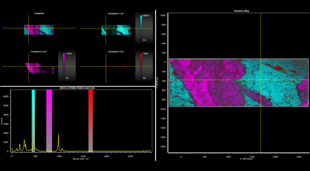
Using an RM5 Raman microscope a sample can be studied by first taking a white light visible image across the gemstone being analysed, and subsequently the Raman map acquired. The sample analysed here was described as sodalite, Figure 1. Sodalite is a strongly blue coloured tectosilicate mineral largely found in Canada and Greenland, but the sample isn’t all blue, so what else is there? When mining for any mineral there will typically be a mix of minerals and the processing of the sample will determine its purity and quality. In this example Raman mapping identified 3 constituents in the sample which can be selected and labelled in the Ramacle® software, Figure 2.

Figure 2: Ramacle® constituent analysis of a gemstone sample.
The Raman map is shown in three colours to represent the three constituents that were identified, Figure 3. The blue areas represent the sodalite that the gemstone the sample was labelled as. The magenta area is orthoclase which is a potassium feldspar material and represents one of the most abundant minerals on Earth. Finally, a small amount of the sample (especially at the top right of the Raman map), shown in red, was identified as nepheline.

Figure 3: Raman spectrum from the 3 constituents found by Raman imaging
Read the full application note to find out more about the use of Raman microscopy in the gemstone industry.
The RM5 Raman Microscope is a compact and fully automated Raman Microscope for analytical and research purposes. The truly confocal design is unique to the market and offers uncompromised spectral resolution, spatial resolution, and sensitivity. To find out how the RM5 can help with your research, please contact us.
If you would like to stay up to date with future news, products and research from Edinburgh Instruments, don’t forget to follow us on social media and sign up to our infrequent eNewsletter via the links below.