Welcome to Edinburgh Instruments monthly blog celebrating our work in Raman, Photoluminescence, and Fluorescence Lifetime Imaging. Every month we will highlight our pick for Map of the Month to show how our spectrometers can be used to reveal all the hidden secrets in your samples.
A laboratory which analyses works of art will typically combine the expertise of historians, restorers, and scientists to determine the authenticity of a work of art. This can be achieved through many techniques, but critically important is that the approach used must be non-destructive as preservation of genuine pieces of art is crucial.
One method to determine whether an art piece, such as a painting, is legitimate or not is through pigment analysis. All paints and inks will contain distinctive pigments, and these pigments can reveal secrets from the painting, such as its age. Different pigment sources have been used throughout different historical periods. When authenticating a painting which is meant to be of a certain age, if modern pigments are identified the analyst can clearly identify the painting as a recent forgery.
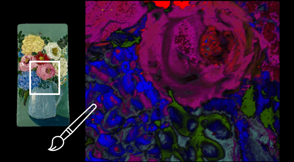
Raman microscopy is one such technique which meets criteria of being non-destructive, whilst also being extremely sensitive to pigments. Raman mapping a painting reveals the different pigments present, and their location within the sample, Figure 1.
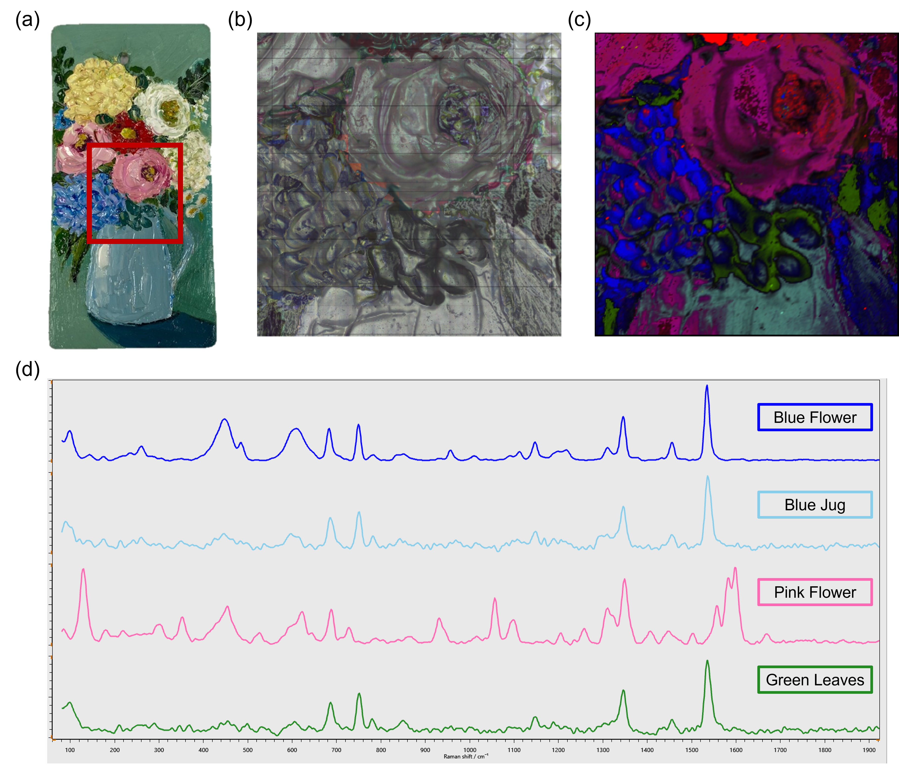
Figure 1: (a) Painting sample (b) white light image of the area to be Raman mapped (c) false coloured Raman map (d) Raman spectra from each colour region of the painting
In this sample we can see the sensitivity of Raman spectroscopy to different pigments, for example, the painting contains two blue regions – the flowers, and the jug holding the flowers. Their spectra are, as expected, very similar, however, there are still clearly some spectral differences visible between the two regions of different blues. More complex data analysis can further be applied to the spectra to reveal more subtle differences between pigments. Combining Raman spectroscopy with a confocal microscope enables Raman mapping across the sample to determine the distribution of these pigments. This can also reveal if colours appearing the same to the human eye were produced by different pigments, which gives art historians more information on the artists techniques and tools available at the time.