Two-dimensional materials have remarkable structural, electronic and optical properties, but understanding how these properties vary across an individual flake requires more than a single measurement.
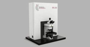
In this Edinburgh Instruments webinar, Angela Flack and Dr Nikesh Patel demonstrate how multimodal microscopy combines Raman spectroscopy, PL and SHG imaging to investigate 2D materials from complementary perspectives. Using the RMS1000 Raman Microscope, the webinar moves from the fundamentals of each technique to live mapping and detailed analysis of molybdenum disulfide and tungsten diselenide samples.
Raman spectroscopy provides a vibrational fingerprint of a 2D material by measuring the frequency shifts produced by inelastic scattering. These vibrational modes are highly sensitive to the material’s structure, enabling Raman mapping to investigate layer number, defects, strain, doping and functionalisation across a sample.
During the live demonstration, Raman mapping of molybdenum disulfide reveals clear differences across individual flakes. The webinar also demonstrates fast mapping, reducing the example Raman map from approximately six minutes with standard mapping to around a minute and a half.
While Raman spectroscopy probes phonons, photoluminescence spectroscopy probes electronic structure and the band gap. Changes in PL emission energy and intensity can reveal variations in layer thickness, defects, strain and interactions with the surrounding environment.
This makes PL imaging particularly valuable for visualising spatial variations across 2D materials. In the molybdenum disulfide example, regions of lower PL intensity are associated with greater layer number, while shifts in PL peak position can indicate potential strain within the flake.
Second harmonic generation takes the analysis a step further by probing crystal symmetry. SHG is a nonlinear optical process that occurs in non-centrosymmetric materials, making its intensity and polarisation highly sensitive to crystal orientation. This enables SHG imaging to investigate crystal stacking, orientation, domains, grain boundaries and defects in 2D materials.
In the live demonstration, SHG mapping reveals distinct domains and grain boundaries that are less apparent in the corresponding Raman and PL maps.
The power of multimodal microscopy becomes particularly clear in the analysis of tungsten diselenide. Raman imaging distinguishes monolayer and multilayer regions, while PL spectral analysis enables regions to be assigned as monolayer, bilayer and trilayer. SHG then adds information about crystal symmetry and stacking.
Together, these complementary techniques connect vibrational structure, electronic properties and crystal symmetry within the same sample, providing researchers with a more comprehensive understanding of their 2D materials. This information can help assess synthesis, identify areas for improvement and support more complete material characterisation.
Watch the full webinar to see Angela Flack and Dr Nikesh Patel showcase Raman, photoluminescence and SHG imaging in action on the RMS1000 Raman Microscope, and discover how combining complementary spectroscopic techniques can uncover more information from 2D materials.