Detection of ionising radiation such as X-rays and γ-rays is important for many applications such as medical imaging, homeland security, and for high-energy physics research. Metal-halide perovskites such as CsPbBr3 have become increasingly popular materials for X-ray detection due to their low production cost, high sensitivity across a wide range of photon energies (200 – 500 keV), and their ability to operate at room temperature.
One of the biggest hurdles for commercialisation of perovskites for this application is achieving high quality bulk crystals as this is the primary factor influencing detector performance. High quality crystals exhibit large vertical diffusion coefficients and long carrier lifetimes which are critical for efficient charge collection.
We highlight work published in Advanced Materials from Professor Samuel Stranks and co-workers at the University of Cambridge.1 In this work, the authors used radioluminescence (RL) spectroscopy to assess the quality of CsPbBr3 crystals for detection of ionising radiation.
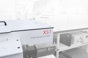
In order to be confident that the RL measurements were a reliable indicator of crystal quality, high-performance, research-grade instrumentation was required. RL measurements were performed on an Edinburgh Instruments FLS1000 Photoluminescence Spectrometer equipped with the XS1 RL chamber (Figure 1). The chamber couples to the FLS1000 via a liquid light guide and is compatible with both continuous wave (CW) and pulsed X-ray sources.

Figure 1: Schematic of an FLS1000 Photoluminescence Spectrometer coupled to an XS1 RL chamber for spectral measurements.
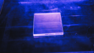
CsPbBr3 single crystal of different qualities were synthesised at different grow rates: low quality (3 mm h-1), high quality A (2 mm h-1), and high quality B (1 mm h-1). Crystal quality was also assessed by their dark counts and γ-ray response as a working detector. RL measurements were performed in an Edinburgh Instruments XS1 coupled to an FLS1000.
Single crystals of varying quality were loaded into the XS1 chamber and were irradiated by an X-ray source with voltage and current set at 60 kV 200 µA, 40 kV 300 µA, or 20 kV 600 µA, for a total of 12 W in each case. X-ray sources provided with the XS1 allow for easy adjustment of voltage and current using software control. Emission was detected on a PMT-980, providing spectral coverage up to 950 nm.
Under irradiation, each of the crystals exhibited emission bands centred at ~550 nm (Figure 2). With the X-ray source operating at 60 kV, 200 µA, the sample with the slowest crystal growth rate, high quality B, produced the most intense signal of the three (Figure 1a). However, at 40 kV, 300 µA, and 20 kV, 600 µA, high quality A crystals produced the most intense signal (Figure 1b, c). While it may have been assumed that slower growth rates would produce higher quality, more strongly emitting crystals, this was not the case. The picture is more complex, showing emission is also dependent on the voltage and current of the source.

Figure 2: RL emission spectra of CsPbBr3 crystals of varying quality measured with X-ray source operating at (a) 60 kV 200 µA, (b) 40 kV 300 µA, and (c) 20 kV 600 µA. Figure reprinted with permission from Stranks et al. 1
This Research Highlight has demonstrated how the FLS1000 coupled to the XS1 may be used to assess the quality of perovskite crystals using RL spectroscopy. The tuneable X-ray source allowed the team to investigate the optimal voltage and current for each sample, providing valuable information about sample quality. Furthermore, the high sensitivity of the FLS1000 allowed the authors to observe weak signals and detect subtle changes in radioluminescence intensity at different source conditions.
The article was published in Advanced Materials and is available at: DOI:10.1002/adma.202512302