Scintillators are materials that convert high-energy ionising radiation, such as X-rays and gamma rays, into UV or visible light. This ability makes them important for applications ranging from medical imaging and baggage scanners to high-energy and particle physics.
Designing a high-performance scintillator requires more than simply observing light emission. Researchers need to understand how efficiently the material converts radiation into light, how rapidly it responds, how sensitive it is, and how stable its performance remains over time. Radioluminescence spectroscopy provides the tools to answer these questions.
In this Edinburgh Instruments webinar, Maria Tesa and Dr Tommy Loan explore the fundamentals of scintillation and demonstrate how spectral and time-resolved radioluminescence measurements can help researchers characterise and optimise scintillator materials.
For inorganic scintillator crystals, scintillation can be considered in three stages: conversion, energy transfer and luminescence. Ionising radiation first generates electrons and positive holes. These charge carriers migrate through the material towards activator or luminescence centres before recombining and releasing UV or visible photons.
Understanding these processes helps explain why properties such as light output and decay time are so important. Light output influences scintillator sensitivity, while decay time determines responsiveness and therefore the timescale of events that a detector can resolve.
A combination of spectral and time-resolved radioluminescence measurements can reveal complementary information about scintillator performance.
Spectral radioluminescence can investigate emission wavelength, defects, detection limits, light yield and linearity. Time-resolved radioluminescence, meanwhile, provides insight into response speed, charge-carrier kinetics, defects and stability.
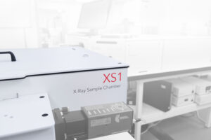
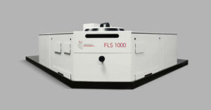
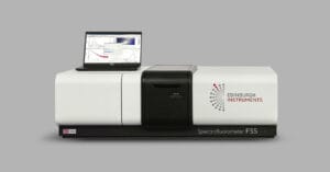
The XS1 X-Ray Radioluminescence Chamber, used with the FLS1000 Photoluminescence Spectrometer or FS5 Spectrofluorometer, supports both steady-state and time-resolved radioluminescence measurements using continuous and pulsed X-ray excitation. The system is designed around a shielded sample chamber, with emitted light coupled to the spectrometer for analysis.
The webinar highlights several applications demonstrating how radioluminescence spectroscopy can guide scintillator development.
In halide perovskite X-ray detectors, researchers compared radioluminescence intensity with gamma-ray detector performance and found that higher-quality crystals exhibited substantially higher radioluminescence intensity. This demonstrates how RL spectroscopy can provide an indicator of crystal quality when screening scintillator materials.
Another study investigated perovskite-metal organic framework (MOF) composites, combining photoluminescence and radioluminescence measurements to assess material stability, sensitivity and response. Continuous radioluminescence measurements examined linearity, detection limits and stability, while pulsed measurements revealed a fast average radioluminescence lifetime of 10 ns for the perovskite-MOF material.
Finally, temperature-dependent radioluminescence measurements of copper(I) cluster scintillators revealed changes associated with defect-related traps and radiative recombination efficiency, illustrating how radioluminescence can move beyond performance testing to uncover the photophysical mechanisms governing scintillator behaviour.
Watch the full webinar to hear Maria Tesa and Dr Tommy Loan explain how continuous and pulsed X-ray radioluminescence spectroscopy can help characterise, compare and optimise scintillator materials for radiation detection and imaging applications.