Welcome to Edinburgh Instruments’ blog celebrating our work in Raman, Photoluminescence, and Fluorescence Lifetime Imaging. Every month, we aim to highlight our pick for Map of the Month to show how our Raman and fluorescence spectrometers can be used to reveal all the hidden secrets in your samples.
 November
 It is officially autumn at Edinburgh Instruments. The clocks have gone back, it is noticeably colder, and we are beginning to do our commutes in the dark with views of fireworks. Perhaps the most noticeable change is the emergence of red, orange, and yellow hues as trees enter dormancy and their leaves begin to change and fall. In this Map of the Month blog, we use photoluminescence microscopy to explore this phenomenon.
Leaves are green because they contain chlorophyll, the chemical contained within chloroplasts in leaf cells that is an essential part of photosynthesis. It absorbs sunlight, giving plants energy vital to convert carbon dioxide into water and sugars. Plants must continuously synthesise chlorophyll to maintain consistent levels, but to do this, they require warm temperatures and sunlight. This is not a problem in the summer months, but it becomes more difficult in the autumn when temperatures and daylight hours drop. At this time of year, chlorophyll production slows, and since decomposing chlorophyll is not replenished, plant leaves begin to lose their green colour. The colouration changes to yellow, orange, and red because the remaining compounds, such as carotenoids and flavonoids, become dominant.
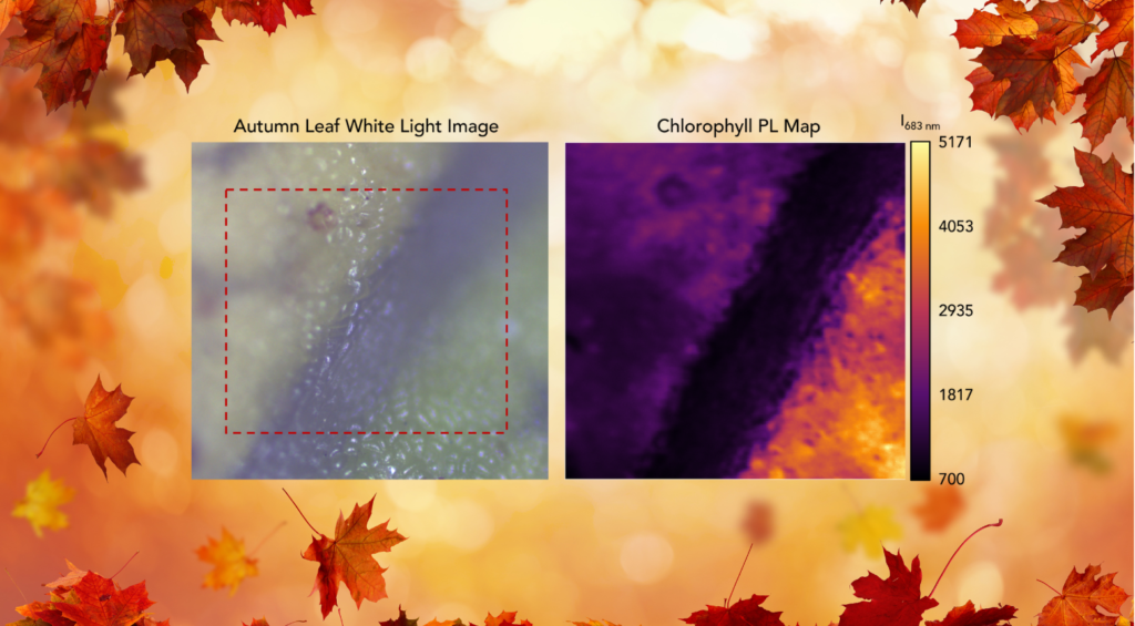
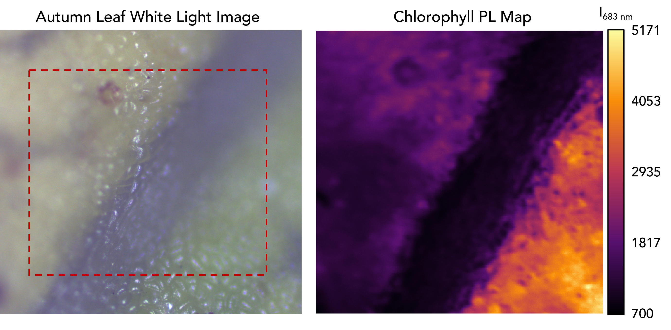
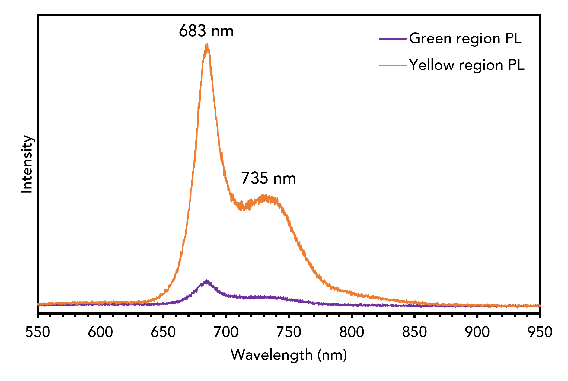
We wondered if we could track this change using spectroscopy, so we picked a fallen leaf from outside our factory and placed it under the RM5 Confocal Raman Microscope for analysis, which is also capable of detecting PL. We identified an area where some remaining green pigmentation mapped its boundary with a yellow-coloured region (above). We used a 532 nm laser and a 300 gr/mm grating to detect PL emissions and saw that the green region on the leaf produced significantly more PL from a peak at 683 nm (the spectra, which are characteristic of chlorophyll, are shown below). The data suggests that the green section of the leaf contains much more chlorophyll than the yellow section.
Enjoyed reading Map of the Month? Stay up to date with our latest MOTM, application notes, blogs and more by subscribing to our newsletter today!